Introduction
All women’s health clinicians know that there are subtle anatomical differences in genital architecture between individuals. But, assumptions that variations in anatomy are infinite, may lead the practitioner to assume that an observed difference is normal when, in fact, it is not. The following is a brief overview of architectural changes that might be seen and must be taken into consideration when making a diagnosis, whether the patient is symptomatic or not.
Significance of abnormal architecture
Any chronic inflammatory condition can produce remarkable scarring of the vulva.1
When scarring alters the architecture of the vulva, the diagnoses of lichen sclerosus, lichen planus, severe atrophy, or previous trauma, must be considered. (Review each of these conditions by searching key words.)
Abnormalities of architecture can develop with symptoms, or develop silently without symptoms. They may appear with changes in skin color, texture, or integrity, or they may not. The skin color may appear to be normal. Treatment of the inflammatory process is imperative to control symptoms if present, prevent the development of pain, and to prevent further architectural changes and scarring.
Abnormalities of vulvar architecture
Changes of the mons and labia majora
Architectural changes of the mons and labia majora are limited to the size of the fat pad, making these areas more prominent in some women. In the postmenopausal woman, atrophy of the fat pads of the labia majora causes reduction in size, although the fat pad of the mons usually remains the same throughout life.
Changes of the labia minora
Hypertrophy of the labia minora may be present at birth, may be produced intentionally in some cultures, may occur from repeated traction associated with sexual practices, or may be idiopathic. Hypertrophied labia minora may be a source of local irritation, problems with genital hygiene, or discomfort with walking, sitting, or sports activities. The hypertrophy may also be a source of discomfort with sexual intercourse. It is essential, however, to distinguish the discomfort of labial irritation from pain on touch in the vestibule (Annotation I, Q-tip test and pain mapping) or irritation from other sources.
Labia minora hypertrophy is generally described as protuberant labial tissue projecting beyond the labia majora. However, there is no consensus among gynecologists, pediatricians, or plastic surgeons regarding the use of objective clinical measurements to confirm the diagnosis.
In an early description of this condition, Friedrich classified labia minora as hypertrophic when the maximal width between the midline and the lateral free edge of the labia minora (when the labia were extended laterally by the examiner) measured greater than 5 cm.2
Others have proposed that the normal width of the labia minora should be up to four cm.3 Some feel that labial reduction should be offered to women (and fully developed adolescents) with persistent symptoms related to this anatomy.4
Variants of labia minora
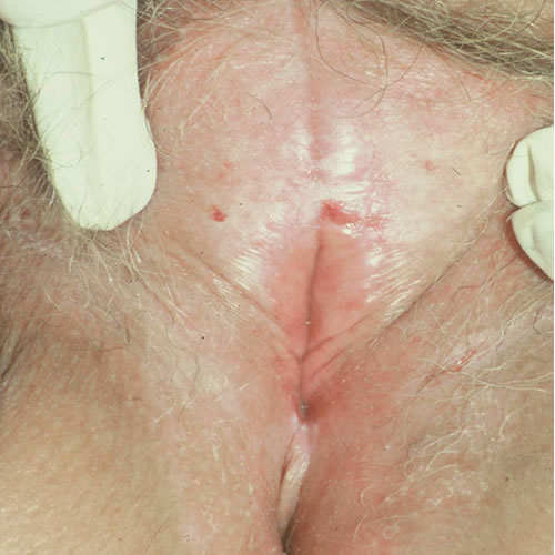
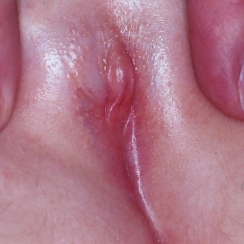
Abnormalities of the labia minora are key indicators of disease, usually dermatosis. Meticulous questioning may uncover a history of itching or irritative symptoms when a woman claims to have been asymptomatic. (Quite frequently, women mention that they have had frequent yeast infections, which, in fact are symptoms relating to the dermatosis.) The inflammation may be active, with whitening, erythema, erosions, ulcers, or fissures, or may be in remission or “burned out,” with abnormal architecture such as labial loss,
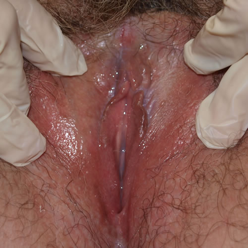
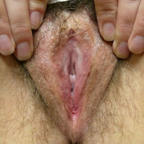
loss of prepuce
or scarring to the glans clitoris with the epithelium normal in color, texture, and integrity.
It is our opinion that women with architectural changes and the appearance of inactive disease should be carefully monitored every six to twelve months, since the natural history of lichen sclerosus and lichen planus is unknown. Further scarring may lead to dyspareunia and/or urinary compromise.
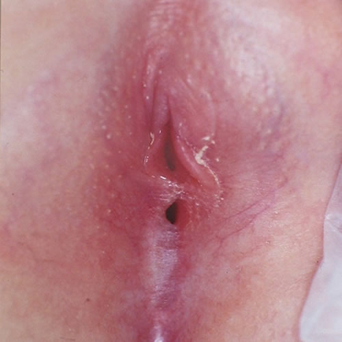
Variants or abnormalities include asymmetry, posterior flattening, and resorption, unilateral or bilateral absence, agglutination of a portion of the labium to the interlabial sulcus,
or “skip areas” where a portion of the labium is missing.
Interlabial sulci will lose their definition if the labia minora are absent.
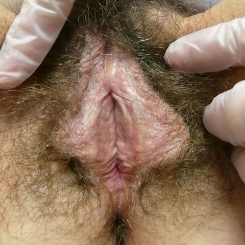
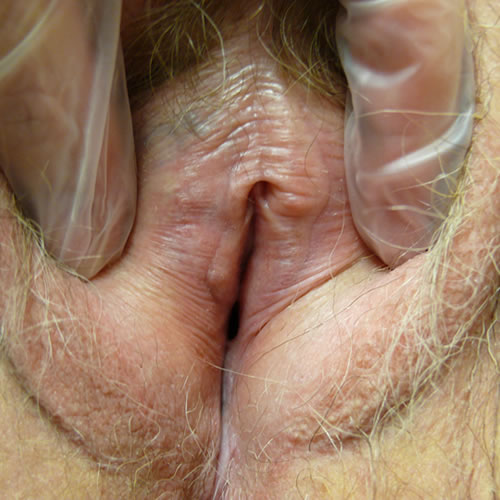
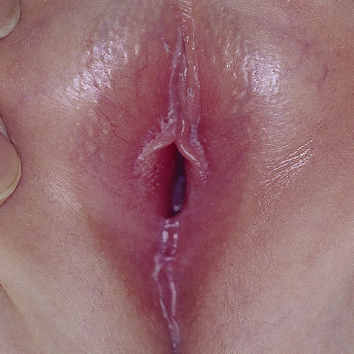
Abnormalities of the prepuce
Abnormalities of the prepuce include loss of the tissue from inflammation so that the glans clitoris is exposed,
thickening and loss of mobility so that the glans is partially visualized, scarring to the glans in one area,

or total fusion with obliteration of the glans.
Lichen sclerosus or lichen planus are well known causes of such scarring,5 but often go unrecognized. The scarring of the prepuce and clitoris are mistakenly given the diagnosis of “phimosis”; without treatment, the inflammatory disease may progress, and scarring may increase. (Lichen sclerosus and Lichen planus).
Abnormalities of the clitoris
Absence of the clitoris, probably as a result of hypoplastic genital tubercles, has been reported at least once.6
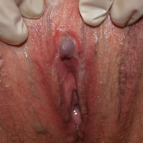
Acquired clitoral enlargement can result from a variety of conditions:
- A clitoral cyst (rare) or neoplasm can enlarge the paraclitoral area without a change in androgens. Recent research documented that tubular eccrine glands form a normal constituent of the sulcus between the glans and the prepuce. These glands may explain rare cysts and some pilonidal sinuses.7 Imaging by ultrasound or MRI is helpful to gain information regarding size and location. Excision is usually necessary by a surgeon skilled with clitoral procedures, often a gynecologic oncologist.
- Exogenous androgen exposure from anabolic steroids, DHEA, or testosterone, may enlarge the glans. Women who use testosterone for therapeutic reasons (treating low libido, averting osteoporosis, or as part of an anti-depressant regimen), may also experience some enlargement of the clitoris although the dosages warranted for these conditions are much lower. Although the treatment of choice for lichen sclerosus is the ultrapotent steroid clobetasol, topical testosterone continues to be prescribed. If there is clitoral enlargement from exogenous androgens, discontinuing the androgen is indicated.
- Endogenous androgen imbalance or excess: In women, mild virilization, the presence of hirsutism, acne, deepening of the voice, frontal (or crown) balding, increased muscle mass, and/or clitoromegaly, indicate the presence of moderate androgen excess arising from stromal hyperthecosis, polycystic ovarian syndrome, ovarian or adrenal neoplasm, or adrenal hyperplasia. An endocrine work-up is necessary.
- Androgen receptor insensitivity (formerly called incomplete testicular feminization) is seen in patients who have an overall female phenotype with female breast development and predominantly female external genitalia except for posterior labial fusion and/or clitoromegaly. Consult with a reproduction endocrinologist is indicated.
Sometimes there may be no obvious or hormonal reason for clitoromegaly after a workup.
Architectural alterations of the vestibule
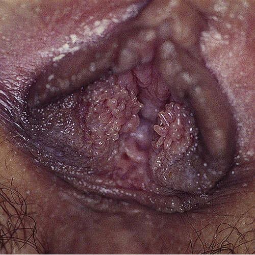
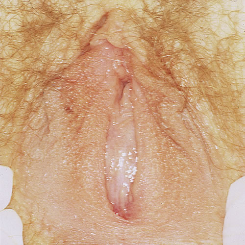
Papillae: These are discrete tubular projections with rounded tips, considered to be normal variants. (Vestibular papillomatosis)
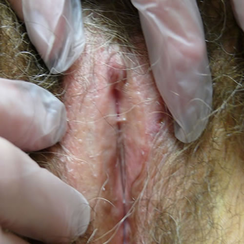
Synechiae: Synechial formation anteriorly or posteriorly, usually from scarring vulvitis such as lichen sclerosus or lichen planus, restricts the vestibule and lessens the introital opening.
The inflammatory condition must be treated, usually with an ultra-potent steroid such as clobetasol applied in a thin film topically, nightly for 30 days and then maintained with twice-weekly use. Then, scarring affecting the introitus may be addressed. Work with progressive sizes of dilators inserted for 20-30 minutes at least three times weekly may gain some added space. (dilator use in Annotation D: Tolerance of genital exam).Physical therapy to the pelvic floor may be necessary to overcome tightening and secondary vaginismus in response to pain.
Surgical division of labial synechiae with cold knife or by laser, in our experience, yields a high rate of recurrence, despite attempts at prevention with such modalities as Inteceed anti-adhesion material, Dermabond coating, and mechanical separation with Vaseline gauze. Some of our patients have had benefit from a post-surgical routine of ongoing control of the dermatosis with an ultrapotent steroid topically twice a week, adequate local estrogen with estradiol cream three times weekly to the vestibule, and topical barrier to adhesion with vaseline or zinc oxide applied three times a day. In addition, 20-30 minutes of dilator use (Annotation D: Patient tolerance for exam, dilators) or intercourse three times weekly maintains patency of the introitus. On-going monitoring every 3-4 months is important. This challenging regimen has to be maintained indefinitely.
Perineoplasty with vaginal advancement may be necessary for comfortable intercourse if division of synechiae and/or dilator therapy fail.
Labial adhesions: Appearing in pediatric or adolescent patients most frequently, the labia can become adherent to each other without the presence of disease.
The blockage is most likely to occur posteriorly, but may also occur anteriorly, in the mid section of the introital opening, or as an almost complete occlusion. If there is no effect on the urinary system, the adhesions may go unnoticed. We have seen a 15 year old who could not insert a tampon due to previously unrecognized labial adhesions (resolved with topical estrogen alone over the course of two months) and a couple of young woman postpartum, whose minor labial tears had not been repaired and had grown together.
Application of estrogen cream on a daily basis has been effective in releasing the adhesions in many of these cases, but may bring on side effects of vaginal bleeding and breast budding in prepubertal girls.8 9 In the case of asymptomatic adhesions in premenarchal girls, the adhesions will usually release on their own as they move into puberty. Topical steroids such as clobetasol or betamethasone 0.05% in a thin film once or twice a day have also been used, with close, monthly follow up, with success. 10
Surgical separation of the adhesions is sometimes required because of urinary retention, pain, or infection.11 Laser has also been used successfully in the treatment of adhesions.
Abnormalities of the hymen
Abnormalities of the hymen include absence of an opening. (Annotation Q, The hymenal ring and the bimanual exam). This may be complete, leading to hematocolpos or partial, leading to problems with tampons or sexual activity.
- Imperforate hymen: Primary amenorrhea is the major symptom, manifesting in puberty with hematocolpos and hematometrium. Cyclic cramping may occur, but many patients are free of symptoms. Diagnosis is made by history and identification of a bulging membrane at the introitus.
Hematocolpos may not occur if there is partial perforation of the hymen, but adolescent women may present with dyspareunia. A cruciate incision into the hymen extending to the 10, 2, and 6 o’clock positions is the treatment of choice.
Imperforate hymen that is present in the neonate can cause urinary retention12 because of fluid from the vagina building up and putting pressure on the urethra. Toddlers or young adolescents can present with urinary retention for the same reason, although this is rare.13 There is one reported case of imperforate hymen associated with other congenital abnormalities (bilateral hydronephrosis, polydactyly and laryngocele) in a female neonate with hydrometrocolpos and urinary retention.14
- Partially ruptured hymen: Women will present with complaint of an unconsummated relationship and dyspareunia. Hymenectomy can be undertaken to help these women achieve penetration with sexual activity. This procedure is optimized with the use of laser, which both cuts and repairs simultaneously. Hymenectomy is NOT the treatment for vaginismus.
- Strictures of the hymenal ring relating to lichen sclerosus or lichen planus: A scarred hymenal ring may be slowly dilated with progressive dilator use with lidocaine; perineoplasty with vaginal advancement may also be required.
Other architectural changes
Other architectural changes may be seen with edema, trauma, and female genital cutting.
References
- Edwards L and Lynch,P. Genital Dermatology Atlas. Philadelphia: Lippincott, Williams & Wilkins, 2004: 97.
- Friedrich EG. Vulvar Disease, 2nd ed. Philadelphia, W.B. Saunders, 1983.
- Rouzier R, Louis-Sylvestre C, Paniel BJ, et al. Hypertrophy of labia minora: experience with 163 reductions. Am J Obstet Gynecol. 2000; 182: 35-40.
- Reddy J, Laufer MR. Hypertrophic labia minora. J Pediatr Adolesc Gynecol. 2010; 23:3-6.
- McPerson T, Cooper S. Vulval lichen sclerosus and lichen planus. Dermatol Ther. 2010; 23: 523-532.
- Falk HC, Hyman AB. Congenital absence of the clitoris: a case report. Obstet Gynecol. 1971;38: 269-271.
- van der Putte SCJ, Sie-go, D. Development and structure of the glandopreputial sulcus of the human clitoris with a special reference to the glandopreputial glands. Anat Rec. 2011; 294:156-64.
- Tebruegge M, Misra I, and Nerminathan V. Is the topical application of oestrogen cream an effective intervention in girls suffering from labial adhesions? Arch Dis Child. 2007 March; 92(3): 268–271.
- Mayoglou L, Dulabon L, et al. Success of Treatment Modalities for Labial Fusion: a retrospective evaluation of topical and surgical treatments. Journal of Pediatric and Adolescent Gynecology. August 2009. Volume 22, Issue 4, 247-250.
- Myers JB, Sorensen CM, Wisner BP, Furness PD 3rd, Passamaneck M, Koyle MA. Betamethasone cream for the treatment of pre-pubertal labial adhesions. J Pediatr Adolesc Gynecol. 2006 Dec;19(6):407-411.
- Mayoglou L, Dulabon L, et al. Success of Treatment Modalities for Labial Fusion: a retrospective evaluation of topical and surgical treatments. Journal of Pediatric and Adolescent Gynecology. August 2009. Volume 22, Issue 4, 247-250.
- Sharifraghdas F, Abdi H, Pakmanesh H, Eslami N. Imperforate hymen and urinary retention in a newborn girl. J Pediatr Adolesc Gynecol. 2009 Feb;22(1):49-51.
- Adali E, Kurdoglu M, Yildizhan R, Kolusari A. An overlooked cause of acute urinary retention in an adolescent girl: a case report. Arch Gynecol Obstet. 2009 May;279(5):701-703. Epub 2008 Sep 6.
- Ozturk H, Yazici B, Kucuk A, Senses DA. Congenital imperforate hymen with bilateral hydronephrosis, polydactyly and laryngocele: a rare neonatal presentation. Fetal Pediatr Pathol. 2010 Jan;29(2):89-94.