Dermatological Terminology
An understanding of basic dermatological terminology1 2 is essential to understand and describe morphologic characteristics in vulvar skin disease. The list of the most important dermatological terms is included here, as well as in Annotation H, where the skin is discussed extensively.
Lesion: a visible or palpable abnormality.
Rash: the word used colloquially to describe numerous or diffuse abnormalities. The lesions within a rash (exanthem) will resemble each other but may vary from being described as macular, papular, vesicular, urticarial, etc. The definitions below are more specific.
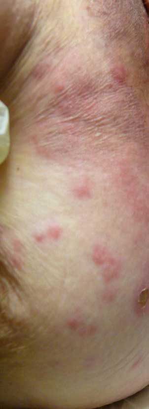
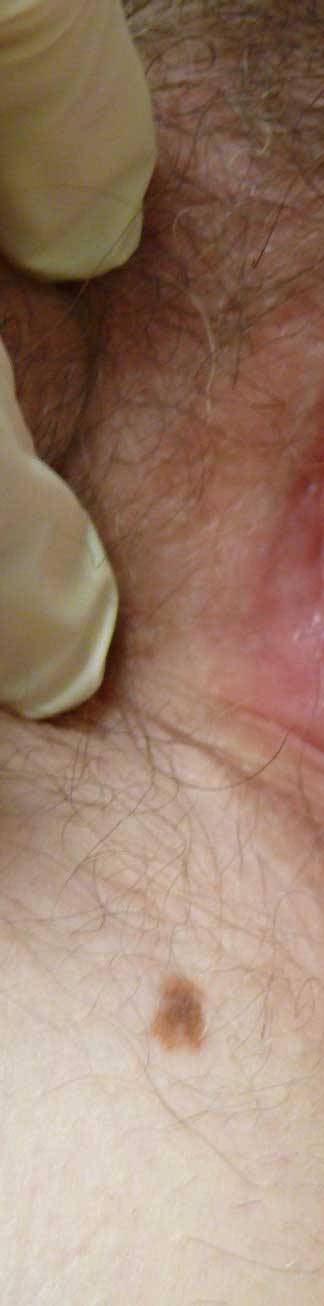
Macule: a small, flat area of color change, (< 1.0 cm in diameter). Not palpable. May be of any color.
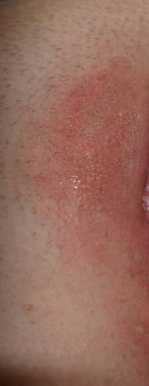
Patch: an extension of a macule in length and width, (>1.0 cm); a non-palpable area of color change.
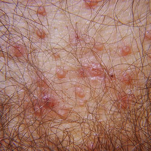
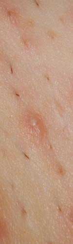
Papule: a small, elevated, palpable lesion (< 1.0 cm in diameter).
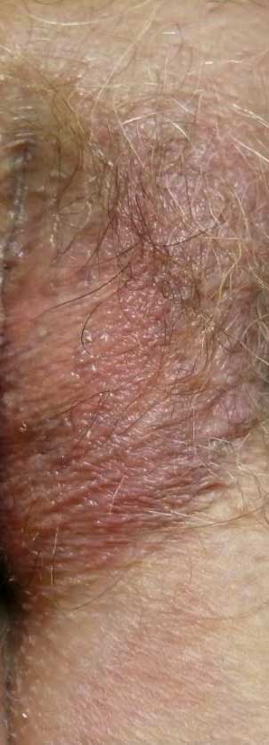
Plaque: an enlargement of a papule in length and width, (>1.0 cm). Elevated, palpable and flat-topped.
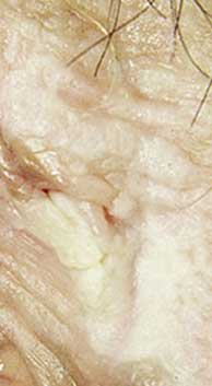
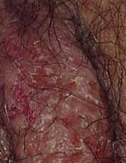
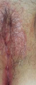
Lichenification: thickening of the skin with increased prominence of skin markings, usually from scratching or rubbing; excoriations may be seen. Scale may or may not be present. Skin may be red, white or skin-colored; the white color is a result of moisture retention in the thickened outer epidermis. This lesion may occur on normal skin or be part of an underlying skin disease.
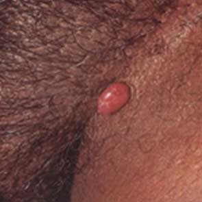
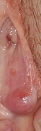
Nodule: a large papule, (>1.0 cm); often hemispherical or poorly marginated; may be located on the surface, within or below the skin; may be cystic or solid.
Cyst: a closed cavity lined by epithelium that contains fluid or semisolid material.
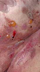
Vesicle: a small, fluid-filled blister (1.0 cm or less in diameter) in which the fluid is clear to slightly cloudy. When the roof of a vesicle has been removed or has disintegrated, an underlying erosion remains visible.
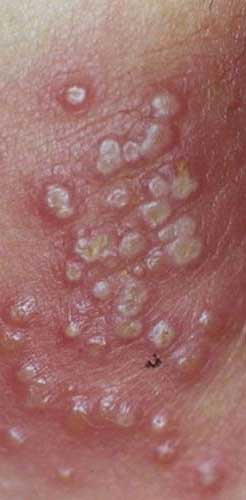
Pustule: a vesicle that is packed with neutrophils and appears to be opaquely white. The fluid in a pustule (pus) is white or yellow.
Bulla: a large vesicle greater than 1.0 cm in diameter, containing clear fluid that is either located in a single compartment or is multiloculated, as a result of coalescence of multiple vesicles or bullae.
Crust: the irregular residue of exudates drying on the skin; implies disruption of the underlying epithelial barrier layer; may be thin or thick. The color will be yellow when made of dried serum, yellow or yellow-green when made of purulent material, or brown, dark red, or brown when made of old blood.
Scale: a hyperproliferative response of the epidermis; grey, white, or silver in color, looking like fine dust but presenting with palpable roughness.
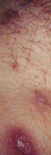
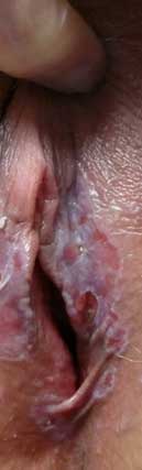
Erosion: a shallow defect affecting the epidermis down to the basement membrane; the dermis is intact. Primary erosions: caused by trauma, usually scratching. These excoriations are linear or angular in shape, without “collarettes.” Secondary erosions: caused by breakdown or removal of a blister roof. These erosions are round and have a collarette of scale encircling the defect.
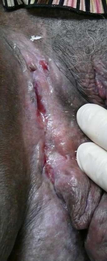
Fissure: a thin, linear erosion of the skin surface.
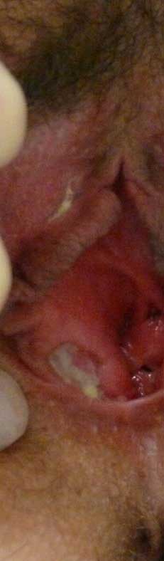
Ulcer: a deeper defect affecting the epidermis and some or all of the dermis.
2006 ISSVD CLASSIFICATION OF VULVAR DERMATOSES: PATHOLOGIC SUBSETS AND THEIR CLINICAL CORRELATES (Table modified from the original)
| Name of finding | Definition | Common clinical correlates |
|---|---|---|
| Spongiosis | Intercellular edema between keratinocytes in the epidermis; keratinocytes may become elongated; often accompanied by exocytosis of lymphocytes and sometimes neutrophils or eosinophils. | Eczematous dermatitis (atopic, contact and allergic) |
| Acanthosis | Diffuse epidermal hyperplasia with increased thickness of the stratum spinosum. | Lichen simplex chronicus, Psoriasis, Reiter’s syndrome |
| Lichenoid pattern | A band-like infiltrate of inflammatory cells (usually lymphocytes) in the superficial dermis, parallel to the epidermis. | Lichen sclerosus, Lichen planus |
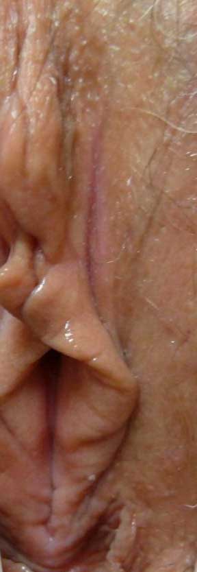
| Dermal sclerosis | Increased collagen with decreased numbers of fibroblasts causing thickening. | Lichen sclerosus |
| Vesiculobullous pattern | Immune deposits in skin may be bands of IgG or IgA at the basement membrane zone, of IgG deposits on the cell surface of keratinocytes. | Bullous pemphigoid, Cicatricial pemphigoid, Pemphigoid gestationis, Pemphigus vulgaris, Pemphigo vegetans |
| Acantholytic pattern | Loss of cohesion between keratinocytes because of dissolution of intercellular connections; may cause an intraepithelial vesicle. Keratinocytes are rounded rather than elongated and lymphocytes do not migrate into the epidermis. | Hailey-Hailey disease, Darier/s disease, Acantholytic dermatosis of the vulvocrural area |
| Granulomatous pattern | Granulomas are conglomerates of monocyte-derived histiocytes (macrophages); sometimes mixed with other inflammatory cells. Groups of these histiocytes may aggregate to form mulinucleated giant cells. Chronic granulomatous diseases are a genetically heterogeneous group of immunodeficiencies producing infectious and non-infectious disease in which phagocytic cells fail to kill organisms that they have engulfed because of defects in a system of enzymes that produce free radicals and other toxic small molecules. | Crohn disease, Melkersson-Rosenthal syndrome |
| Vasculopathic pattern | Non-inflammatory purpura: extravasation of RBCs into dermis. Narrowing or obliteration of small vessel lumina. Inflammation of blood vessel wall. | Aphthous ulcers, Behcet disease, Plasma cell vulvitis (vulvitis plasmacellularis) |
References
- Lynch PJ, Moyal-Barracho M, Scurry J, Stockdale C. 2011 ISSVD Terminology and Classification of Vulvar Dermatological Disorders: An Approach to Clinical Diagnosis. Journal of Lower Genital Tract Disease, Vol 16, 4, 2012, 339-344.
- A. Nast, C.E.M. Griffiths, R. Hay, W. Sterry, J.L. Bolognia, The 2016 International League of Dermatological Societies’ revised glossary for the description of cutaneous lesions, British Journal of Dermatology, Volume 174, Issue 6, 1 June 2016, Pages 1351–1358, https://doi.org/10.1111/bjd.14419