Introduction
Eczema and atopic dermatitis are synonyms for morphologically and histologically identical skin conditions.1 Atopy refers to a genetic predisposition for hypersensitive allergic IgE reactions to common environmental antigens,2 with allergic rhinitis (hay fever), asthma, and/or atopic dermatitis that looks exactly like eczema. “Eczematous” refers to the characteristic appearance of the skin (see below). Atopic individuals may be more likely to develop eczematous skin reactions to common exposures that would not bother the non-atopic person. (Contact dermatitis in the Atlas). We thus emphasize the important component of vulvovaginal care that stresses the elimination of possible irritants: tight clothing, daily panty liners, panty hose, feminine care products, harsh soaps, or perfumes. At the same time, eczema may occur in those who have no family or personal history of atopy, and atopic dermatitis is rare in the vulvar area.
Another term clear to dermatologists but unknown to lay people is the term lichen simplex chronicus (LSC). Lichen simplex chronicus is closely linked with itching and may occur anywhere on the skin, including on the vulva. Itching causes the individual to scratch or rub, exacerbating the itch sensation. The vicious cycle of itching-scratching-rubbing-itching ends up causing the specific morphological condition of LSC: erythema that may occur in plaques of varying depths, in pustules, excoriations, rough scaling that appears shiny rather than flaking, areas of hypopigmentation or hyperpigmentation, especially in people with naturally dark skin. The final hallmark sign of LSC is lichenification: thickening of the skin with the distinctive pattern of normal skin “hatch-markings” being exaggerated and deepened when compared with adjacent, non-affected skin. In addition, because of scratching, hair may be absent or broken. Lichen simplex chronicus may occur because of itching associated with another skin disease such as lichen sclerosus or candidiasis, or may develop as de novo itching. (See Lichen simplex chronicus in the Atlas).
Histologically, eczema, dermatitis, atopic dermatitis, and lichen simplex chronicus appear the same. A semantic assumption is made by many dermatologists that lichen simplex chronicus is the descriptive term that encompasses localized eczematous conditions in the vulva, atopic dermatitis the term that encompasses generalized conditions, and that the clinician’s job is to come to understand what might have precipitated the condition and treat both the LSC and the originating condition. In the third edition of their excellent textbook, Genital Dermatology Atlas and Manual, Libby Edwards and Peter Lynch make this statement: “Given the lack of concensus regarding the diagnostic criteria for atopic dermatitis and in view of the fact that these eczematous eruptions share the presence of the “itch-scratch cycle,” I believe it is appropriate to use the term atopic dermatitis (for the multifocal form) and LSC (for the localized for) of the disease regardless of whether or not the individual has other clinical evidence of atopy. Moreover, since genital eczematous involvement very often occurs in the absence of eczematous disease elsewhere, it will generally be referred to as LSC for the purpose of this chapter.”3
Epidemiology
It is impossible to realistically determine prevalence of the eczematous vulvar conditions because they overlap with other conditions and because the defining terminology also overlaps. However, eczematous dermatidides are very common and affect up to 20% of children and 10% of adults globally.4 More than 31 million Americans have some form of eczema.5 6 Atopic dermatitis, the generalized type of eczema that appears on the non-vulvar skin, occurs in 15% of the population in the Western world.7
Etiology
The etiology of eczematous dermatitis includes several elements. Among these are genetic risk factors, skin barrier dysfunction, immune dysregulation, altered skin microbiome, and environmental triggers of inflammation. Skin barrier dysfunction, in particular, is recognized as a key factor in the initiation and progression of eczematous dermatitis as it leads to vulnerability to infection, allergens, and irriatnts, frequently culminating in pruritis.8
Symptoms and clinical features
The patient presents with complaint of itching and dry skin. In some cases, she will have put off making an appointment to the point that itching has been partially replaced by pain from scratching or by burning from urine or fabrics touching the affected areas. Assuming a yeast infection, she may mention vaginal discharge, as well, which may not be relevant to her condition. Nighttime pruritis is common. The patient may not be aware of scratching while asleep. Heat and sweat, as well as friction from physical activity or tight clothing may provoke flares of itching.9
Clinical features include:
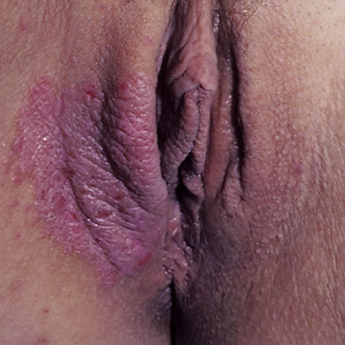
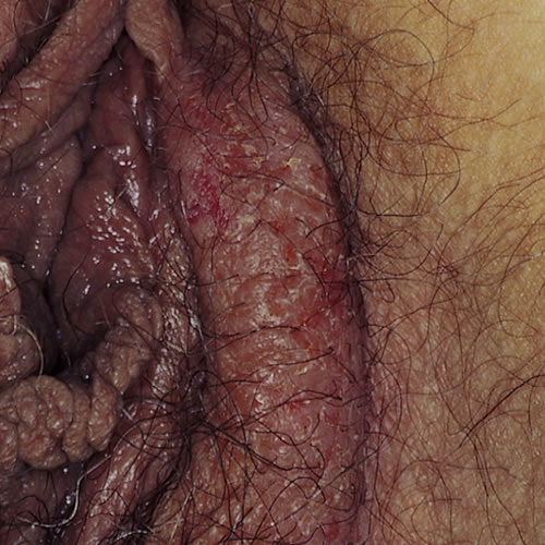
Erythema in varying degrees, from color barely distinguishable from that of surrounding tissue to deep red. In dark-skinned people, erythema may present as hyperpigmentation, darkening, rather than redness.
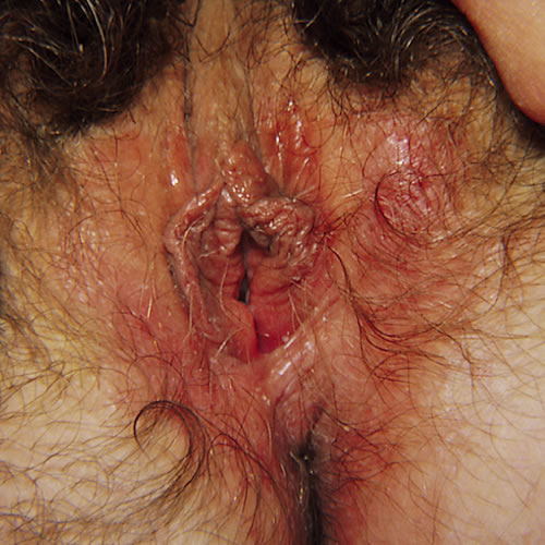
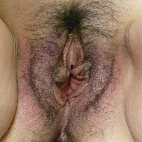
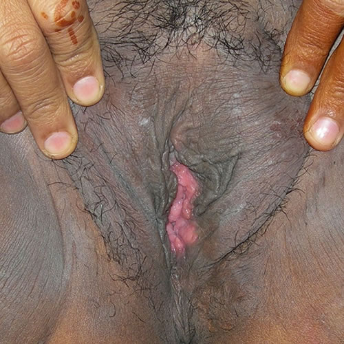
Red plaques with poorly marginated borders. Subacute and chronic eczematous dermatitis presents as dry, scaly, erythematous to hyperpigmented plaques. Overlying scale which may be seen as tiny white flakes or may present as barely perceptible roughness of the skin, or as shiny, tight appearing skin.
Epithelial disruption in the form of excoriations, fissuring, weeping, crusting or yellow scale.10
Lichenification in the form of thickened skin, exaggerated skin markings and lichen type scale which is shiny rather than flaking. This is the end result of the itch-scratch-itch cycle known as lichen simplex chronicus. See Lichen simplex chronicus
Pathology/Laboratory Findings
Because eczematous conditions can be caused by instigators such as Candida, contact with allergens or irritants, and lichen simplex chronicus can also be stimulated by skin response to underlying dermatoses such as lichen sclerosus, careful detective work includes wet mount and microscopy, yeast cultures and, if the diagnosis is not clear, biopsy. Acanthosis and spongiosis are most frequently seen on biopsy. If superimposed bacterial infection is suspected (usually if there are weeping wounds), bacterial culture should be done.
Diagnosis
Diagnosis is accomplished by a combination of scrupulous history taking,11 clinical observations, and histopathology. Full-body cutaneous examination may reveal clues to assist in narrowing the differential diagnosis, including eczematous changes on other skin surfaces or stigmata of atopy such as xerosis (abnormally dry skin or mucous membranes), keratosis pilaris (dry, rough patches and tiny bumps, often on the upper arms, thighs, cheeks or buttocks), follicular accentuation (bumps around hair follicles), Dennie-Morgan lines (folds seen under the eyes), or hyperlinear palms.
Differential Diagnosis
Differential diagnosis includes psoriasis, seborrheic dermatitis, contact dermatitis, and eczematous Candidiasis.
Treatment and management
Treatment for all the eczematous vulvar skin conditions is the same.
- Identify contributing factors such as yeast infection and treat them. Culture for yeast even if it is not seen under the microscope. Because the use of topical steroids, the primary treatment for eczematous vulvar skin conditions, may make women more susceptible to Candidiasis, consider treatment for yeast even if it is not seen. (See table below).
- Encourage the wearing of loose, comfortable, non-binding clothing. Examine life style issues that may have brought the condition on. Eliminate irritants or allergens and treat the skin with soothing warm water soaks, patting dry gently.
- Cool gel packs, bags of frozen peas wrapped in soft cloth, or frozen water bottles, similarly softened, may be soothing.
- Application of a thin film of petrolatum can be helpful in sealing in moisture and making the patient more comfortable.
- Scratching must stop! Reduce itching with topical Lidocaine 5% applied up to six times a day as needed to quell the itching. (Warn that Lidocaine may burn initially, but will numb the skin after about 60 seconds).
- Reduce night time scratching with hydroxyzine 10 to 25 to 50 mg orally at bedtime if the patient desires it. Benadryl is sometimes helpful as an alternative that patients may be more familiar with.
- Patients may have developed a habit of scratching that is hard to break, especially because the scratching brings temporary relief or pleasure. Helping them become more aware of the behavior is a first step. Adjunctive treatment with SSRIs (SSRIs in treatment plans) may be of use.
- Steroids, the dose and route of medication dictated by the severity of the condition, are extremely important in the resolution of eczematous skin conditions. In general, topical steroid ointments range in potency from low to mid to super potent. Low grade itching and skin that is not too inflamed can be treated with low potency steroid ointment. More extensive discomfort and skin disruption can be treated with the higher dose ointments. For extensive disorders or those that are unresponsive to topical treatment, intramuscular or oral steroids can be helpful. Intralesional steroids are used in the case of very thick plaques or localized but recalcitrant disease. See table below.
- After the symptoms are controlled, the frequency of application and the potency of the cortisone can be gradually reduced, as indicated, moving from the mid potencies down to a mild steroid such as 1% to 2.5% hydrocortisone or desonide 0.05% ointment. Avoid creams.
See table below.
Treatment of Eczematous Vulvar Conditions
References
- Brown S, Reynolds NJ. Atopic and non-atopic eczema. BMJ. 2006 Mar 11;332(7541):584-8. doi: 10.1136/bmj.332.7541.584. PMID: 16528081; PMCID: PMC1397720.
- Edwards L and Lynch PJ. Genital Dermatology Atlas, third edition. Wolters Kluwer Health and Lippincott Williams and Wilkins, 2018. p 67.
- Edwards L and Lynch PJ. Genital Dermatology Atlas, third edition. Wolters Kluwer Health and Lippincott Williams and Wilkins, 2018. p 67.
- Ständer S. Atopic Dermatitis. N Engl J Med. 2021 Mar 25;384(12):1136-1143. doi: 10.1056/NEJMra2023911. PMID: 33761208.
- Hanifin JM, Reed ML; Eczema Prevalence and Impact Working Group. A population-based survey of eczema prevalence in the United States. Dermatitis. 2007 Jun;18(2):82-91. doi: 10.2310/6620.2007.06034. PMID: 17498413.
- Silverberg JI, Hanifin JM. Adult eczema prevalence and associations with asthma and other health and demographic factors: a US population-based study. J Allergy Clin Immunol. 2013 Nov;132(5):1132-8. doi: 10.1016/j.jaci.2013.08.031. Epub 2013 Oct 4. PMID: 24094544.
- Edwards L and Lynch PJ. Genital Dermatology Atlas, third edition. Wolters Kluwer Health and Lippincott Williams and Wilkins, 2018. p 67.
- Ständer S. Atopic Dermatitis. N Engl J Med. 2021 Mar 25;384(12):1136-1143. doi: 10.1056/NEJMra2023911. PMID: 33761208.
- Ständer S. Atopic Dermatitis. N Engl J Med. 2021 Mar 25;384(12):1136-1143. doi: 10.1056/NEJMra2023911. PMID: 33761208.
- Fisher, BK and Margesson LJ. Genital Skin Disorders: diagnosis and treatment. Mosby, 1998. 154-158.
- Woelber L, Prieske K, Mendling W, Schmalfeldt B, Tietz HJ, Jaeger A. Vulvar pruritus-Causes, Diagnosis and Therapeutic Approach. Dtsch Arztebl Int. 2020 Feb 21;116(8):126-133. doi: 10.3238/arztebl.2020.0126. PMID: 32181734; PMCID: PMC7081372.