Introduction
Contact dermatitis1 is a general term used to describe inflammation of the skin resulting from an external agent that acts as either an irritant or an allergen to produce a rash that can be acute (or subacute), or chronic. Vulvar and other anogenital skin is uniquely at risk for contact dermatitis because the skin barrier is more likely to be compromised by moisture from urine, sweat, and discharge, as well as by friction, occlusion, and low estrogen states, all of which increase the likelihood of penetration or absorption of irritants or allergans.2
It is important to work with the patient to determine if the agent is an irritant or an allergen. Find out precisely what products the patient uses and how often. The history is paramount.
Epidemiology
Irritant contact dermatitis of the non-vulvar skin accounts for 80% of occupational skin disease. Its prevalence reveals that it can be a source of discomfort to anyone exposed to an irritant or toxin.3 The prevalence of irritant contact dermatitis in the genital area is unknown, although one study placed it at 20%,4 a number that rings true for us in our practice.
Allergic contact dermatitis is common in general, accounting for 7% of occupational illnesses. Non-occupational allergic contact dermatitis is reported to be three times more common than the occupational.5 However, in the vulvar area, allergic reactions are found less frequently than irritant reactions. Allergic contact dermatitis is uncommon in young children and people older than 70 years of age.6
Patients with pre-existing dermatitides and atopic diathesis (atopic dermatitis, allergic rhinitis, and asthma) are at higher risk for developing vulvar contact dermatitis.7
Etiology, symptoms, and clinical features
Irritant contact dermatitis: acute and chronic
Acute irritant contact dermatitis is produced very rapidly in reaction to exposure to an irritant and results in erythema, edema, and sometimes blistering. The reaction may take place within minutes or up to 24 hours after the exposure. Because of the rapid onset of reaction, the patient is usually more aware of what might have caused it. Because blisters do not last long on non-keratinized skin, erosions or ulcers may form on the inner aspects of the labia minora or the vestibule when they become unroofed. If the affected skin is keratinized (outer structures of the vulva), the blisters may last longer and be seen by the clinician. Patients complain of burning, stinging, or pain.
The irritants that cause an acute reaction are usually medications applied topically to the vulva to treat warts or Candida, laser treatments, or harsh chemicals that the women themselves have chosen to use to remove perceived discharge, odor, or uncleanliness.8
Chronic irritant contact dermatitis can be more difficult to diagnose than the acute irritant reaction because symptoms of irritation, pain, soreness, or rawness can develop slowly. Itching can be a symptom in atopic individuals. (See Atopy below). Often, the skin barrier has been disrupted in such a way that it is either too wet or too dry.9 Chronic irritant contact dermatitis is usually caused by prolonged exposure to milder forms of irritants such as soap, water, urine, or feces, topical products such as feminine sprays or washes, bases of some gels or creams, or low-grade yeast infections. Ordinary exposure to water, soaps, toilet paper, pads, tampons, etc, has not been shown to cause irritant reactions.10
In both acute and chronic irritant contact dermatitis, the reaction is caused by the physically irritating or caustic effect of the substance.
Allergic contact dermatitis
An allergy is a hypersensitivity disorder of the immune system. Allergic reactions occur when a person’s immune system reacts to normally harmless substances in the environment as if they were pathogens. These reactions are acquired, predictable, and relatively rapid. While allergic reactions of the respiratory system result in asthma or rhinitis, allergic reactions in the vulva present with symptoms of itching. These are cell-mediated, delayed type (type IV) hypersensitivity reactions and they usually occur 48 hours to days after exposure.
The most common cause of allergic contact dermatitis in the vulvar area is exposure to a perfume, preservative, or a medication that has been applied to the skin or mucosa. Exposure to poison ivy or other plant in nature likely to cause reactions is also possible.
Semen allergy may cause local reactions, and, once the patient is sensitized, may even cause anaphylaxis. (Annotation J: Lifestyle issues for semen allergy). This is true of latex allergy, as well.
Atopy
Atopy is a predisposition to having allergic reactions. Atopic individuals are those who are more likely than others to have eczema (atopic dermatitis), hay fever (allergic rhinitis or conjunctivitis), or allergic asthma, as well as food allergies. Atopic reactions are thought to be IgE-mediated, localized hypersensitivity reactions with a possible hereditary component. Although atopic dermatitis is rare in the vulvar area, a tendency to be “atopic” is thought to possibly predispose some women to irritant or allergic contact dermatitis.
A personal or family history of allergies, eczema and asthma represents atopy and is significant information.
List of Allergens and Irritants in Vulvar Contact Dermatitis
- Allergens
Antibiotics — neomycin, bacitracin
Anesthetics — benzocaine, dibucaine
Preservatives — parabens, imidazolidinyl urea, propylene glycol
Perfumes — balsam of Peru
Moisturizers — lanolin
Plants — poison ivy
Rubber — gloves, condoms, and diaphragms
Spermicides
Seminal plasma fluid - Irritants
Soaps and detergents
Fabric softeners
Feminine hygiene products — pads, diapers Wipes, feminine deodorant sprays
Physically abrasive contactants — face cloths
Sponges
Sweat, urine, feces, etc
Hot water bottles — Thermally damaging
Podophyllin and similar products
Trichloracetic and bichloroacetic acid
Laser and other hair removal processes
Symptoms and clinical features
The patient’s main complaints are of itching, burning, and irritation that may be sudden in onset or gradual and cumulative depending on the etiology. The patient might or might not be aware of the product or exposure that precipitated the problem. The symptoms may worsen with heat and moisture.
In irritant contact dermatitis, there may be a rapid reaction to an applied substance: usually irritation, burning, or rawness; (itching is less frequently found than in the allergic reactions). There is often a history of repeated use of the product. This is particularly true of the soap and feminine hygiene products.
Some patients are obsessive cleansers of the vulvar area and seriously irritate their skin in the process. Elderly women may use normally harmless products that, when combined with napkins for incontinence or panty hose or girdles, can produce a reaction equivalent to a chronic irritant “diaper dermatitis” in infants.
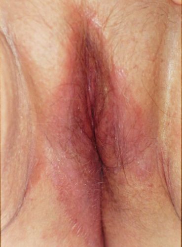
The patient has poorly demarcated, slightly scaly, erythematous patches or slightly elevated plaques without edema. The erythema is in darker red or brownish tones rather than the bright red of the allergic reaction. The vulvar skin can appear dry, glazed, fissured, and chapped looking. If the irritant stimulated itching and scratching, the appearance of the skin can resemble atopic dermatitis or lichen simplex chronicus.
In allergic contact dermatitis, the development of itching, swelling, and irritation may pinpoint the precise cause, such as exposure to latex in a condom or a particular cream or ointment used. If the condition is severe, there may be pain and burning on micturition.
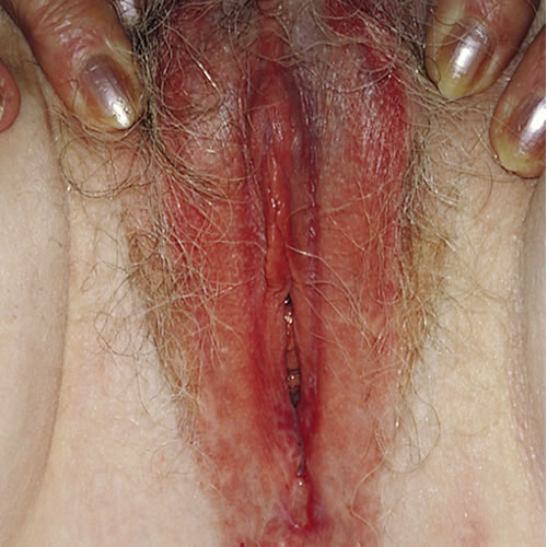
Acute involvement of the vulvar area can cause swelling, vesiculation, erythema, and weeping. Scratching may cause secondarily infected excoriations with crusting. The pattern may be subacute with some degree of erythema, swelling, erosion, and crustiness, or chronic with lichenification, induration, erythema, and either hypopigmentation or hyperpigmentation.
Patients often have poorly marginated, bright red, erythematous patches and plaques, often on the labia majora. Scale is not seen as often as in other types of dermatitis. Urticaria does not usually occur on the vulva.11 12
Diagnosis
History of atopy, products used and clinical pattern help determine the diagnosis.
Pathology/Laboratory Findings
Biopsy may be helpful in confirming the diagnosis if necessary, but will usually simply show “acanthosis and spongiosis: irritant or allergic reaction.”
To investigate allergic contact dermatitis, patch testing can be done by a dermatologist. (Allergists often do prick testing that is not as extensive as the North American Series of dermatologic patch tests.) Irritant reactions are more frequently found than allergic reactions.
Differential diagnosis
Atopic dermatitis, candidiasis, psoriasis, intertrigo, and tinea or, rarely: extra-mammary Paget disease, Hailey-Hailey disease, Darier disease.
Treatment/management
Rule out yeast with yeast culture even if no yeast is seen under the microscope. Consider the possibility of secondary bacterial infection and culture if indicated.
The most important treatment is to stop exposure to the allergen or irritant. For extensive allergic reactions, a dermatologist or allergist should be consulted.
Local care for weeping, irritated skin and severe pruritus includes warm or cool sitz baths or cold compresses and ventilated cotton clothing to relieve the symptoms. Topical petrolatum is a soothing barrier.
Topical corticosteroids that can be prescribed include:
- Betamethasone-17-valerate 0.1% ointment applied twice a day in a thin film until improved
OR
- Triamcinolone acetonide 0.1% ointment twice a day applied in a thin film until improved
If the symptoms are severe, consider:
- Prednisone 0.5 to 1 mg/kg/day orally decreased over 14 to 21 days
- Triamcinolone acetonide 40 mg/mL (Kenalog 40) 1 mg/kg IM single dose
- Antihistamine (as a sedative) — hydroxyzine 25 to 75 mg orally at bedtime
Note: After the symptoms are controlled, the frequency of application and the potency of the cortisone can be gradually reduced, as indicated, moving from the mid potencies down to a mild steroid such as 1% to 2.5% hydrocortisone or desonide 0.05% ointment. Avoid creams.
See (Treatment Plan) for more details.
References
- Fisher BK, Margesson, LJ. Genital Skin Disorders: Diagnosis and Treatment. Mosby, Inc., 1998. 155-156.
- Woodruff CM, Trivedi MK, Botto N, Kornik R. Allergic Contact Dermatitis of the Vulva. Dermatitis. 2018 Sep/Oct;29(5):233-243. doi: 10.1097/DER.0000000000000339. PMID: 30179968.
- Wolff K and Johnson RA. Fitzpatrick’s Color Atlas & Synopsis of Clinical Dermatology, sixth edition. McGraw Hill, 2009. 21.
- Genital pruritus and the eczematous diseases in Genital Dermatology Atlas, 2nd edition. Wolters Kluwer/Lippincott Williams & Wilkins. Philadelphia, 2011. 38.
- Wolff K and Johnson RA. Fitzpatrick’s Color Atlas & Synopsis of Clinical Dermatology, sixth edition. McGraw Hill, 2009. 26.
- Wolff K and Johnson RA. Fitzpatrick’s Color Atlas & Synopsis of Clinical Dermatology, sixth edition. McGraw Hill, 2009. 26.
- Corazza, M.; Toni, G.; Zedde, P.; Schettini, N.; Borghi, A. Contact Dermatitis of the Vulva. Allergies. 2021, 1, 206-215. https://doi.org/10.3390/allergies1040019
- Corazza, M.; Toni, G.; Zedde, P.; Schettini, N.; Borghi, A. Contact Dermatitis of the Vulva. Allergies. 2021, 1, 206-215. https://doi.org/10.3390/allergies1040019
- Farage MA, Miller KW, Berardesca E, Maibach HI. Incontinence in the aged: contact dermatitis and other cutaneous consequences. Contact Dermatitis. 2007 Oct;57(4):211-7. doi: 10.1111/j.1600-0536.2007.01199.x. PMID: 17868212.
- Corazza, M.; Toni, G.; Zedde, P.; Schettini, N.; Borghi, A. Contact Dermatitis of the Vulva. Allergies. 2021, 1, 206-215. https://doi.org/10.3390/allergies1040019
- Lynch PJ. Genital pruritus and the eczematous diseases in Genital Dermatology Atlas, 2nd edition. Wolters Kluwer/Lippincott Williams & Wilkins. Philadelphia, 2011. 38.
- Lynch PJ. Genital pruritus and the eczematous diseases in Genital Dermatology Atlas, 2nd edition. Wolters Kluwer/Lippincott Williams & Wilkins. Philadelphia, 2011. 41-43.