Introduction
Lymphatic malformation is a benign proliferation of the lymphatic vessels, either congenital (lymphangioma circumscripta [LC] or deeper cavernous lymphangioma) or acquired (lymphangiectasia [AL]). Congenital lesions mature as patients mature and their external genital organs develop until the lesions become apparent in adulthood.1 LC can occur anywhere in the body, but vulvar presentation is uncommon.
Epidemiology
Lymphatic malformation accounts for 4% of all vascular malformations and 25% of all benign vascular tumors.2 The uncommon vulvar presentation of LC, either congenital or acquired, includes approximately 31 cases. 3
Etiology
LC results from a hamartomatous malformation of lymphatic vessels.4 AL occurs when lymphatics are obstructed after surgery and radiotherapy for a malignant tumor, long-standing Crohn disease, hidradenitis and, rarely, tuberculosis.5
Symptoms and clinical features
Lesions may go unnoticed for many years. They present as asymptomatic swellings. These lesions can itch and burn. In the vulvar area, they may rupture and discharge lymph, causing local irritation.
In both LC and AL, the lesions appear like a mass of “frog’s eggs” with groups of 1-5 mm translucent or hemorrhagic vesicles on skin-colored bases or a reddish-brown base.
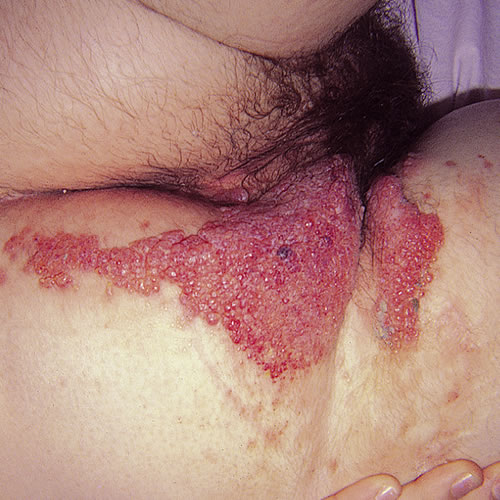
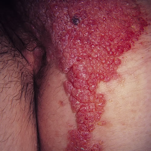
There may be several groups of these vesicles. Hyperkeratosis may give the vesicles a verrucous appearance, leading to the misdiagnosis of viral warts.6 Leakage of lymph can be a problem but confirms the diagnosis.
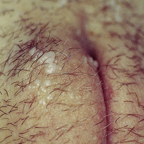
Oozing vesicles and papules should bring into mind the possibility of lymphangiesctasia. Deeper cavernous lymphangioma may not have any superficial surface changes, presenting as swelling. We have followed a case of LC thought to be a Bartholin cyst; pathology from excision confirmed lymphatic etiology. The cyst recurred with development of another on the opposite side; sclerotherapy and excision have been successful treatment.
Diagnosis
Diagnosis is made on clinical observation and histopathology from biopsy.
Pathology/Laboratory Findings
Histologically, AL is indistinguishable from LC.7 Lymphangiomas are composed of dilated endothelial-lined spaces, and the lumens may contain lymphoid cells, red blood cells, or both. There may be overlying hyperkeratosis and acanthosis of the squamous epithelium. Valvular structures may be seen in the dilated channels, distinguishing the lesion from hemangioma.8
Differential diagnosis
Condylomata, Bartholin cyst, angiosarcoma, dermatitis herpetiformis, herpes simplex, herpes zoster, malignant melanoma, metastatic carcinoma of the skin, neurofibromatosis
Treatment
No treatment is needed for asymptomatic lesions.9
Treatment for AL describing manual lymphatic drainage, remedial exercises, skin care and compressive garments is described in Vulvar Edema (Vulvar Edema in the Atlas).
In the past, complete excision has been difficult because of the difficulty in delineating the extent and depth of the lesions. There has been a high recurrence rate. Recently, MRI has been helpful in defining the degree of involvement and the anatomy of the lesion. Excisional surgery and laser treatment are now major treatment modalities in addition to cryotherapy, electrocoagulation, and sclerosing agent injection.10 Some experts consider excisional sugery the only plausible choice in the therapy of advanced disease.11
Consultation with an expert from a vascular or lymphedema service located in major medical centers is helpful.
References
- Vlastos AT, Malpica A, Follen MA. Lymphangioma circumscriptum of the vulva: a review of the literature. Obstet Gynecol. 2003; 101(5, Part 1):946-954.
- Colange E, Wilson JE. Vascular tumors, tumors and tumour like conditions of blood vessels and lymphatics. In: Elder De, Elenitsas-Johnson BL Jr, Murphy GF, eds. Levers histopathology of skin, 9th ed. Philadelphia, Lippincott Williams and Wilkins, 2005, 1046-1047.
- Cecchi R, Bartoli L, et al. Lymphangioma circumscripta of the vulva of late onset. Act Dermatol Venereol 1995; 75(1):79-80.
- Mehta V, Nayak S, Monga P, Rao R. Extensive congenital vulvar lymphangioma mimicking genital warts. Indian J Dermatol. 2010; 55(1):121-122.
- Esquivias G, Miranda-Romero A, Cuadrado Valles C, et al. Lymphangioma curcumscriptum of the vulva. Cutis. 2001; 67(3):229-232.
- Jappe U, Zimmermann B, Kahle, et al. Lymphangioma circumscriptum of the vulva following surgical and rediological therapy of cervical cancer. Sex Transm Dis. 2002;29(9):533-535.
- Okur MI, Kose R, Yildirum AM, Cobanoglu B. Lymphangiectasia of the vula accompanying congenital lymphedema. Dermatol Online J. 2009; 15(4): http://dermatology.cdlib.org/1504/letters/lymphiectasia/kose/html
- Heller DS and Wallach RC, editors. Vulvar Disease: a clinicopathological approach. Informa Healthcare.2007, 151.
- Vlastos AT, Malpica A, Follen MA. Lymphangioma circumscriptum of the vulva: a review of the literature. Obstet Gynecol. 2003; 101(5, Part 1):946-954.
- Ahn SJ, Chang SE, Choi JH, et al. A case of unresectable lymphagioma circumscriptum of the vulva successfully treated with OK-432 in childhood. J Am Acad Dermatol. 2006; 55(5 Suppl):S106-107.
- Ahn SJ, Chang SE, Choi JH, et al. A case of unresectable lymphagioma circumscriptum of the vulva successfully treated with OK-432 in childhood. J Am Acad Dermatol. 2006; 55(5 Suppl):S106-107.