Introduction
Vulvar cancer is relatively rare, accounting for 3-5% of gynecological and 0.3% of all cancers, with an expectation of 6470 new cases and 1,670 deaths in 2023.1 2 3 Vaginal cancers are more common, and are closely associated with HPV infection of the cervix.4 For the past 10 years, the incidence of vulvar cancer has been increasing by an average of 0.6% per year. Patients with vulvar cancer generally have a poor prognosis due to the high recurrence rate and tendency to metastasize, with a 5-year survival chance of 27%–60%.5
Vulvar squamous cell carcinoma (VSCC), accounting for 85 to 90% of cases of vulvar cancer,6 is the most common type of malignant vulvar tumor, followed by melanoma (5-10%), basal cell carcinoma (2-4%), Bartholin gland adenocarcinoma, sarcoma, and vulvar Paget disease (sporadic).7 8
With the advent of the HPV vaccine, the rate of cervical cancer decreased by 65% in women in their 20’s between 2012 and 2019, an impressive and optimistic statistic that gives hope for some types of vulvar and vaginal squamous cell carcinoma, as well.9
VSCC presents as both in situ vulvar intraepithelial neoplasia and invasive squamous cell carcinoma.
Epidemiology
The annual rate of VSCC in the USA is about 1 to 2 per 100,000.10 The incidence of VSCC has risen, partly because of the increase in HPV infections especially those affecting younger women,11 but is expected to decrease, again with the increased use of HPV vaccine in young people.12
The average age at diagnosis of vulvar cancer (as specific statistics for VSCC are lacking) remains 65-74, with the incidence per 100,000 by race/ethnicity as follows:
Incidence of vulvar cancer per 100,000 in 202313
All races 2.5
Hispanic 1.8
Non-Hispanic American Indian/Alaska Native 2.3
Non-Hispanic Asian/Pacific Islander 1.0
Non-Hispanic Black 1.9
Non-Hispanic White 3.0
Etiology
VSCC develops in two ways: in the presence of HPV infection (HPV+) or in the absence of HPV (HPV-). Some studies term these two pathways “HPV related” or “associated” and “HPV independent.” In both cases, VSCC is caused by a precursor lesion. In the case of HPV associated (HPV +) conditions, the precursor is similar to those that cause intraepithelial lesions anywhere else in the anogenital tract (most commonly with HPV 16, 18, and 33, but also with 31, 45, 52 and 58 strains) and is identified as vulvar high grade squamous intraepithelial lesion (VHSIL), previously known as usual type vulvar intraepithelial neoplasia or uVIN.
The precursor to HPV independent (HPV-) VSCC is known to occur in the setting of chronic inflammatory vulvar conditions such as lichen sclerosus (LS) 14 and is known as dVIN or differentiated vulvar intraepithelial neoplasia. Many studies show a poorer prognosis and higher rate of recurrence for those with HPV- lesions. 15 16
In distinguishing between the two conditions, molecular studies have shown that specific gene mutations can be associated with each. The World Health Association mandates the use of p16 immunohistochemistry in differentiating between HPV+ and HPV- VSCC as this testing shows good correlation with HPV+ effects, making it a surrogate marker for HPV.17 P53, a caretaker gene encoded in TP53, is an important guardian of the human genome. TP53 mutations frequently show up in HPV- lesions, so measurement of p53 can be an indicator for this condition.
Mutations in TP53 can lead to loss of its protector capacity, promoting wild-type p53. Present in many cancers, TP53 gene mutations have been seen in 30-80% of HPV- VSCC, 18 leading to recommendations for immunohistological measurement of the p53 protein, as well as p16.20
There can be complications in identifying and properly categorizing these lesions both morphologically and histopathologically. Some studies have identified areas of cross over, where in the setting of one diagnosis, the other entity was seen and the additional possibility of “hit and run oncogenesis” caused by a “hit” of HPV followed by clearing of the virus in the setting of HPV- lesions. Or, HPV may be present as an “innocent bystander.” These variations can obscure the correct diagnosis. 21
Some researchers are also suggesting a possible third pathway to HPV- VSCC. Identification of two new precursor lesions: vulvar acanthosis with altered differentiation (VAAD) and differentiated exophytic vulvar intraepithelial lesion (DE-VIL) are thought to be associated with HPV- disease.22
Precursor lesions to VSCC are covered in HSIL and dVIN.
Symptoms and clinical features
Precursor lesions can be very subtle and hard to see and HPV+ and HPV- lesions can be indistinguishable. From velvety grayish or skin-colored macules or papules, early VHSIL in HPV+ lesions can progress to flat topped plaques varying in diameter from 0.5-3 cm, usually surrounded by normal appearing tissue, from the vestibule out onto keratinizing skin.23 These plaques, occurring in younger women, can enlarge and become congruent and are usually not symptomatic. Color varies from skin-colored to white, red, pink, black, or brown.24
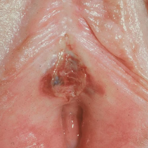
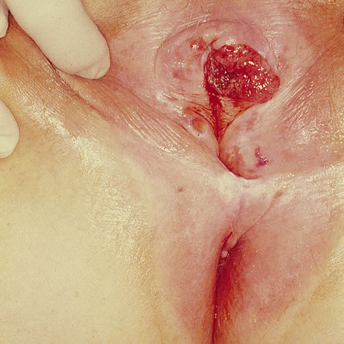
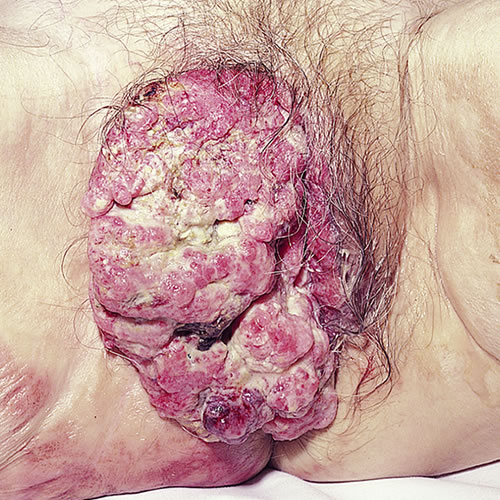
Vulvar dVIN in HPV- VSCC presents differently. Occurring in older women, usually with a history of lichen sclerosus, (and, more rarely, lichen planus), these lesions are seen as unifocal plaques or nodules measuring 2 to 5 cm in size, accompanied by crusting, erosion, and scaling. They may be red, white, brown, appearing in the vestibule or the interlabial sulci with associated itching, pain, and sometimes bleeding, developing ulcerations if not treated.25
Lichen sclerosus itself, especially if not treated, can occur with thickened white plaques, hyperpigmented areas, and redness, obscuring any other suspicious lesions. Regularly scheduled follow up visits of patients with LS is imperative.
Diagnosis
Diagnosis is made with incisional or excisional biopsy preferably with adjunctive immunohistological testing for p16, p53, and in further exploration, HPV ISH (HPV In Situ Hybridization) testing. HPV testing of the cervix, vagina, and any suspicious lesions is also recommended prior to biopsy.
Colposcopic examination may be of additional value.
Laboratory findings
On histopathology, HPV+ HSIL commonly has a warty or basaloid morphology whereas HPV- dVIN shows differentiated epithelium with abnormal maturation and basal atypia.26
It is advisable to take biopsies from different parts of the lesion or from multiple abnormal looking sites if necessary to avoid sampling error or diverse conditions being present at the same time. (See Anno G for biopsy)
Utilization of molecular and epigenetic information for diagnosis, prognosis determination, and treatment planning is rapidly growing in other types of cancer.27 This growing field will inevitably influence care. In the meantime, instruction from dermatopathologists at your own institution about necessary test materials is advised.
Differential diagnosis
Differential diagnosis includes VIN precursors, anogenital warts, seborrheic keratoses, psoriasis, Paget disease, amelanotic melanoma, chancroid, Crohn disease, and metastatic carcinoma.
Treatment/management
Refer to a gynecologic oncologist. The tumor has to be well assessed, staged, and then specific treatment planned for tumor removal and follow up. Please see this excellent 2023 article for the most up to date information on assessing and treating vulvar squamous cell carcinoma: European Society of Gynaecological Oncology Guidelines for the Management of Patients with Vulvar Cancer – Update 2023.
References
- https://seer.cancer.gov/statfacts/html/vulva.html
- Tessier-Cloutier B, Pors J, Thompson E, Ho J, Prentice L, McConechy M, Aguirre-Hernandez R, Miller R, Leung S, Proctor L, McAlpine JN, Huntsman DG, Gilks CB, Hoang LN. Molecular characterization of invasive and in situ squamous neoplasia of the vulva and implications for morphologic diagnosis and outcome. Mod Pathol. 2021 Feb;34(2):508-518. doi: 10.1038/s41379-020-00651-3. Epub 2020 Aug 13. PMID: 32792599.
- Siegel RL, Miller KD, and Jemal A, Cancer statistics, 2020. CA Cancer J Clin, 2020 70(1): p. 7–30
- Siegel RL, Miller KD, Wagle NS, Jemal A. Cancer statistics, 2023. CA Cancer J Clin 2023; 73:17.
- Wei S, Li L, Yi T, Su L, Gao Q, Wu L, OuYang Z. Epidemiologic characteristics and a prognostic nomogram for patients with vulvar cancer: results from the Surveillance, Epidemiology, and End Results (SEER) program in the United States, 1975 to 2016. J Gynecol Oncol. 2023 Nov;34(6):e81. doi: 10.3802/jgo.2023.34.e81. Epub 2023 Jul 5. PMID: 37477104; PMCID: PMC10627757.
- Weinberg D, Gomez-Martinez RA. Vulvar cancer. Obstet Gynecol Clin North Am. 2019;46:125–35.
- Berek JS and Karam A. Vulvar cancer: Epidemiology, diagnosis, histopathology, and treatment. UpToDate.com, Wolters Kluwer. Nov, 2023. Accessed 1/6/24
- Wei S, Li L, Yi T, Su L, Gao Q, Wu L, OuYang Z. Epidemiologic characteristics and a prognostic nomogram for patients with vulvar cancer: results from the Surveillance, Epidemiology, and End Results (SEER) program in the United States, 1975 to 2016. J Gynecol Oncol. 2023 Nov;34(6):e81. doi: 10.3802/jgo.2023.34.e81. Epub 2023 Jul 5. PMID: 37477104; PMCID: PMC10627757.
- Siegel RL, Miller KD, Wagle NS, Jemal A. Cancer statistics, 2023. CA Cancer J Clin. 2023 Jan;73(1):17-48. doi: 10.3322/caac.21763. PMID: 36633525.
- Weinberg D, Gomez-Martinez RA. Vulvar cancer. Obstet Gynecol Clin North Am. 2019;46:125–35.
- Joura E.A., Lösch A., Haider-Angeler M.G., Breitenecker G., Leodolter S. Trends in vulvar neoplasia. Increasing incidence of vulvar intraepithelial neoplasia and squamous cell carcinoma of the vulva in young women. J. Reprod. Med. 2000;45:613–615. [PubMed] [Google Scholar] [Ref list]
- Dehlendorff C, Baandrup L, Kjaer SK. HPV vaccination before 17 years of age reduces the risk of vulvar and vaginal HSIL+ based on real-world data. Real-World Effectiveness of Human Papillomavirus Vaccination Against Vulvovaginal High-Grade Precancerous Lesions and Cancers. J Natl Cancer Inst. 2021 Jul 1;113(7):869-874. doi: 10.1093/jnci/djaa209. PMID: 33377930; PMCID: PMC8246827.
- https://seer.cancer.gov/statfacts/html/vulva.html
- Griesinger LM, Walline H, Wang GY, Lorenzatti Hiles G, Welch KC, Haefner HK, Lieberman RW, Skala SL. Expanding the Morphologic, Immunohistochemical, and HPV Genotypic Features of High-grade Squamous Intraepithelial Lesions of the Vulva With Morphology Mimicking Differentiated Vulvar Intraepithelial Neoplasia and/or Lichen Sclerosus. Int J Gynecol Pathol. 2021 May 1;40(3):205-213. doi: 10.1097/PGP.0000000000000708. PMID: 32925443; PMCID: PMC7960553.
- Griesinger LM, Walline H, Wang GY, Lorenzatti Hiles G, Welch KC, Haefner HK, Lieberman RW, Skala SL. Expanding the Morphologic, Immunohistochemical, and HPV Genotypic Features of High-grade Squamous Intraepithelial Lesions of the Vulva with Morphology Mimicking Differentiated Vulvar Intraepithelial Neoplasia and/or Lichen Sclerosus. Int J Gynecol Pathol. 2021 May 1;40(3):205-213. doi: 10.1097/PGP.0000000000000708. PMID: 32925443; PMCID: PMC7960553.
- Tessier-Cloutier, B., Pors, J., Thompson, E. et al. Molecular characterization of invasive and in situ squamous neoplasia of the vulva and implications for morphologic diagnosis and outcome. Mod Pathol 34, 508–518 (2021). https://doi.org/10.1038/s41379-020-00651-3
- Oonk MHM, Planchamp F, Baldwin P, Mahner S, Mirza MR, Fischerová D, Creutzberg CL, Guillot E, Garganese G, Lax S, Redondo A, Sturdza A, Taylor A, Ulrikh E, Vandecaveye V, van der Zee A, Wölber L, Zach D, Zannoni GF, Zapardiel I. European Society of Gynaecological Oncology Guidelines for the Management of Patients with Vulvar Cancer – Update 2023. Int J Gynecol Cancer. 2023 Jul 3;33(7):1023-1043. doi: 10.1136/ijgc-2023-004486. PMID: 37369376; PMCID: PMC10359596.
- Xing D, Fadare O. Molecular events in the pathogenesis of vulvar squamous cell carcinoma. Semin Diagn Pathol. 2021 Jan;38(1):50-61. doi: 10.1053/j.semdp.2020.09.010. Epub 2020 Sep 25. PMID: 33032902; PMCID: PMC7749059.
- Tessier-Cloutier, B., Pors, J., Thompson, E. et al. Molecular characterization of invasive and in situ squamous neoplasia of the vulva and implications for morphologic diagnosis and outcome. Mod Pathol 34, 508–518 (2021). https://doi.org/10.1038/s41379-020-00651-3[/efn_note] These markers of disease may also serve as indicators for prognosis. The presence of HPV+ and wild type TP53 tumors may show a better prognosis than those found with TP53 mutations. .19 Oonk MHM, Planchamp F, Baldwin P, Mahner S, Mirza MR, Fischerová D, Creutzberg CL, Guillot E, Garganese G, Lax S, Redondo A, Sturdza A, Taylor A, Ulrikh E, Vandecaveye V, van der Zee A, Wölber L, Zach D, Zannoni GF, Zapardiel I. European Society of Gynaecological Oncology Guidelines for the Management of Patients with Vulvar Cancer – Update 2023. Int J Gynecol Cancer. 2023 Jul 3;33(7):1023-1043. doi: 10.1136/ijgc-2023-004486. PMID: 37369376; PMCID: PMC10359596.
- Griesinger LM, Walline H, Wang GY, Lorenzatti Hiles G, Welch KC, Haefner HK, Lieberman RW, Skala SL. Expanding the Morphologic, Immunohistochemical, and HPV Genotypic Features of High-grade Squamous Intraepithelial Lesions of the Vulva with Morphology Mimicking Differentiated Vulvar Intraepithelial Neoplasia and/or Lichen Sclerosus. Int J Gynecol Pathol. 2021 May 1;40(3):205-213. doi: 10.1097/PGP.0000000000000708. PMID: 32925443; PMCID: PMC7960553.
- Tessier-Cloutier, B., Pors, J., Thompson, E. et al. Molecular characterization of invasive and in situ squamous neoplasia of the vulva and implications for morphologic diagnosis and outcome. Mod Pathol 34, 508–518 (2021). https://doi.org/10.1038/s41379-020-00651-3
- Edwards L and Lynch P. Genital Dermatology Atlas and Manual, third edition. 2018. Wolters Kluwer. P 59.
- Edwards L and Lynch P. Genital Dermatology Atlas and Manual, third edition. 2018. Wolters Kluwer. P 59.
- Edwards L and Lynch P. Genital Dermatology Atlas and Manual, third edition. 2018. Wolters Kluwer. P 59.
- Griesinger LM, Walline H, Wang GY, Lorenzatti Hiles G, Welch KC, Haefner HK, Lieberman RW, Skala SL. Expanding the Morphologic, Immunohistochemical, and HPV Genotypic Features of High-grade Squamous Intraepithelial Lesions of the Vulva With Morphology Mimicking Differentiated Vulvar Intraepithelial Neoplasia and/or Lichen Sclerosus. Int J Gynecol Pathol. 2021 May 1;40(3):205-213. doi: 10.1097/PGP.0000000000000708. PMID: 32925443; PMCID: PMC7960553.
- Tessier-Cloutier B, Pors J, Thompson E, Ho J, Prentice L, McConechy M, Aguirre-Hernandez R, Miller R, Leung S, Proctor L, McAlpine JN, Huntsman DG, Gilks CB, Hoang LN. Molecular characterization of invasive and in situ squamous neoplasia of the vulva and implications for morphologic diagnosis and outcome. Mod Pathol. 2021 Feb;34(2):508-518. doi: 10.1038/s41379-020-00651-3. Epub 2020 Aug 13. PMID: 32792599.