Introduction
Pemphigus vulgaris1 is a rare, autoimmune, vesiculobullous disease of the skin and mucous membranes that, without treatment, is potentially fatal.
Epidemiology
The mean age at onset is between 50 and 60 years.2 Pemphigus vulgaris occurs worldwide, but its prevalence depends on geographic location and ethnicity. Incidence rates between 0.1 and 0.5 per 100,000 people per year have been reported, but higher rates are seen in certain populations. Individuals with Jewish ancestry, particularly Ashkenazi, and people who live in India, Southeast Europe, and the Middle East have the greatest risk.3 In Jerusalem, the incidence is estimated at 16 to 32 per million per year, while in France, the incidence is 1.7 per million per year.4
Etiology
The etiology involves an autoimmune process induced by a combination of endogenous and exogenous factors, resulting in production of IgG autoantibodies directed against the intercellular substance and membrane antigens of epithelial cells, the adhesion molecule desmoglein (Dsg).5 The resulting inflammation causes separation (acantholysis) of the epidermal cells as their intercellular bridges disintegrate in the suprabasal layers of both the epidermis and the mucous membrane areas.
Symptoms and clinical features
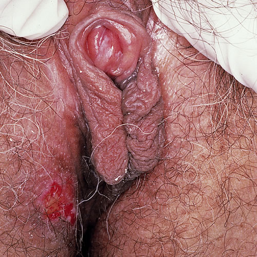
Almost all patients with pemphigus vulgaris have mucous membrane erosions and over 50% will have skin blistering and erosions.6 The mouth is generally the first area involved, and patients complain of burning, soreness, irritation, and difficulty chewing. The vulva is affected in about 50% of cases, with complaints of soreness, burning, and irritation, as with any erosive vulvar condition.7 The lesions in the vulva may be asymptomatic.
Recurrent vulvar ulcers and erosions on the inner labia minora and vulvar vestibule are observed.
Intact blisters are more likely to be noted on keratinized skin. If bullae occur, especially in mucosal areas, the fragile blisters frequently rupture, so that erosions are seen more commonly. Although the bullae eventually heal without scars, long-term disease can result in loss of vulvar tissue, clitoris, and labia; the vagina can be involved, with subsequent scarring. Oral involvement is usually earliest and most symptomatic. Skin involvement elsewhere on the face and body causes open erosions and crusting.
Diagnosis
Clinical picture and biopsy with appropriate immunofluorescent testing defines the diagnosis.
Pathology/Laboratory Findings
Biopsy is essential for the diagnosis and histological exam of a 4mm sample from the edge of a blister or erosion will show the basal cells attached to the basement membrane but not to each other or to cells above them (characterized as a “row of tombstones”). Cells from within the blister will show intraepithelial acantholysis. Direct immunofluorescence of samples of more normal appearing skin adjacent to the blister will show deposits of IgG between the epidermal cells. These samples need to be preserved in Michel’s medium rather than formalin.
If the cervix is involved, pap smears may show changes that are indistinguishable from cervical intraepithelial neoplasia.8 9 10
Differential diagnosis
Differential diagnosis includes mucous membrane pemphigoid (MMP) (cicatricial pemphigoid), lichen planus, erythema multiforme, bullous pemphigoid, drug reaction, and paraneoplastic pemphigus.
Treatment/management
This condition is managed by a dermatologist. With proper management, patients can really do quite well, but without treatment, this is a life threatening condition. Many patients need long-term surveillance. Due to the rarity of the condition, there is a lack of consensus about the best approach to management, however, the use of systemic glucocorticoids has proven to be life saving in many instances. The current risk of death is about 5% and is usually due to drug-related complications including sepsis.
Nonspecific care for the acute swollen vulva includes:
- Gentle cleansing with Cetaphil cleanser
- Avoidance of harsh, irritating soaps
- Wearing loose, ventilated clothing
- For painful open areas, compresses or sitz baths using Burow’s 1:40 solution are recommended to the patient.
Also suggested is the use of a bland barrier ointment as for pemphigus.
Specific treatment for the acute swollen vulva includes the following progressive therapeutic options:
- Topical superpotent steroid (halobetasol or clobetasol) 0.05% ointment applied topically twice a day; can also be used with a Premarin applicator for intravaginal involvement (2 g vaginally at bedtime).
- Corticosteroids—1 mg/kg/day prednisone, decreasing by 5 to 10 mg/day/month; at 20 mg/day of prednisone, add a steroid sparing agent (below), gradually decreasing the prednisone by 5 to 2.5 mg/day/month.
Choices for steroid sparing agents include methotrexate 15-25 mg/week, cyclophosphamide 1.5 to 2.5 mg/kg/day, azathoprine 1.5 to 2.5 mg/kg/day, mycophenolate mofetil 2-3 g/day, and cyclosporine 5 mg/kg/day. Plasma exchange and extracorporeal photophoresis have also been used. IVIG and rituximab are newer, very promising treatment options for pemphigus vulgaris.11 Menses can be suppressed (as in MMP).
References
- Fisher BK, Margesson, LJ. Genital Skin Disorders: Diagnosis and Treatment. Mosby, Inc., 1998. 181-182.
- Amagai, M. “Pemphigus.” Dermatology, edited by Jean Bolognia, Elsevier, Limited. 2018.
- Hertl M and Cassian S. Pathogenesis, clinical manifestations, and diagnosis of pemphigus. UpToDate, Wolters Kluwer, 2022.
- Amagai, M. “Pemphigus.” Dermatology, edited by Jean Bolognia, Elsevier, Limited. 2018.
- Akhyani M, Chams-Davatchi C, Naraghi Z, et al. Cervicovaginal involvement in pemphigus vulgaris: a clinical study of 77 cases. Br J Dermatol. 2008; 158: 478.
- Amagai, M. “Pemphigus.” Dermatology, edited by Jean Bolognia, Elsevier, Limited. 2018.
- Akhyani M, Chams-Davatchi C, Naraghi Z, et al. Cervicovaginal involvement in pemphigus vulgaris: a clinical study of 77 cases. Br J Dermatol. 2008; 158: 478.
- Kanwar AJ, Dhar S. Factors responsible for death in patients with pemphigus. J Dermatol. 1994; 21:655.
- Martin LK, Werth VP, Villaneuva EV, Murrell DF. A systematic review of randomized controlled trials for pemphigus vulgaris and pemphigus foliaceous. J Am Acad Dermatol. 2011; 64:903.
- Amagai, M. “Pemphigus.” Dermatology, edited by Jean Bolognia, Elsevier, Limited. 2018.
- Amagai, M. “Pemphigus.” Dermatology, edited by Jean Bolognia, Elsevier, Limited. 2018.