Introduction
Genital warts are caused by the human papillomavirus (HPV).1 They are among the most common sexually transmitted infectious diseases. Synonyms for HPV include venereal warts, anogenital warts, condylomata acuminata, and HPV infection.
Cervical HPV is discussed in Annotation M Speculum Exam and Cervix.
Vaginal HPV is discussed in Annotation P Obtain pH, Examine Vaginal discharge.
Epidemiology
HPV infects mostly young adults between the ages of 16 and 25 years. The incidence of HPV has risen dramatically in the last twenty years and is increased in those who are HIV positive or who have other sexually transmitted infections.
Etiology
A double-strand DNA papovavirus, HPV has over 70 subtypes. The most common infections (90% of cases) are caused by types 6 and 11. These low risk subtypes are mostly associated with benign condyloma and low grade intraepithelial neoplasia whereas subtypes 16 and 18 can be associated with squamous cell carcinoma. The incubation period can be from three weeks to eight months.
Human Papillomavirus Type and Clinical Disease
| Clinical Disease | HPV Type |
|---|---|
| Common warts | 1,2,4,7 |
| Anogenital warts | 6,11,16,18 Less common 31,33,35,51,52 |
| Genital intraepithelial neoplasia Vulvar + Penile intraepithelial neoplasia Bowen’s disease Bowenoid papulosis |
16,18,31,33,35,51,52* |
| Carcinoma Squamous cell carcinoma (cervix) |
16, 18 |
*Only 3 to 10% of anogenital warts are caused by these types. Only 0.1% of the population develops anogenital malignancy. Types 16 and 18 are strongly associated with squamous cell carcinoma of the cervix.
Transmission of the virus can be sexual or nonsexual. The majority of cases in adults are sexually transmitted. Children may acquire genital warts from infected caregivers. Of children with anogenital warts, 30% to 40% of them have been abused and this must always be ruled out.
Symptoms and clinical features
Usually there is a history of exposure, weeks to months previously, to a sexual partner who has venereal warts. There may be a history of prior warts. Some patients are immunosuppressed. The lesions are usually asymptomatic. If very large and exophytic, they can be traumatized, fissured, and sore.
Initial pinhead papules develop into filiform papules that are often symmetric and on apposing skin surfaces. The lesions of condylomata acuminata can be small or large, skin-colored to reddish, and may grow into cauliflower-like clusters.
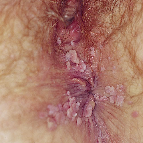
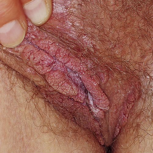
Their surfaces can be smooth or velvety. They are located around the vaginal introitus and perianal area most commonly. A less common clinical pattern is that of papules or plaques, single or multiple, skin-colored or hyperpigmented.
Note: Fifty percent (50%) of women with genital warts will have associated HPV of the cervix.
Diagnosis
Diagnosis is clinical but can be confirmed by biopsy. Rule out neoplasia in patients with HPV using colposcopy and biopsy if indicated by history or by atypical or suspicious features of the lesions, such as ulcerations. In addition, it is important to rule out other associated sexually transmitted diseases. Pap smear guidelines need to be followed. LINK ASCCP
Pathology/Laboratory Findings
Vinegar (acetic acid 5%) applied for 5 to 10 minutes to keratinized skin and for 1 minute to nonkeratinized mucous membrane highlights the HPV lesions (acetowhitening). Excision of a lesion allows for pathological identification if in doubt. Biopsy or excision should be undertaken in the case of atypical, pigmented or suspicious lesions or those that do not resolve after three treatments.
Differential diagnosis
Differential diagnosis includes molluscum contagiosum, lichen planus, enlarged sebaceous glands, condyloma lata (caused by secondary syphilis and appearing flat and velvety), and vulvar intraepithelial neoplasia. Papillomatosis, a normal vulvar variant, can be distinguished from condyloma accuminatum by noting the usual symmetry of the lesions and the way each papilloma arises from a single base. PICTURE (Vestibular papillomatosis).
Treatment/management
No treatment is guaranteed to cure genital warts, and patients should understand and must accept the unpredictability of response to treatment. Risk of recurrence is 30-70%. Up to 30% of cases spontaneously regress. Most treatment modalities are destructive in nature. Choice of treatment can be based primarily on the number and location of lesions
Indications for treatment of genital warts include the following:
- Manage symptoms of itching or burning
- Prevent spread
- Treat disfiguring lesions
- Return or preserve normal function, for example, stop post-coital bleeding or dyspareunia
The following treatments are listed in order of preference.
Provider applied methods:
- Cryotherapy — A cotton-tipped applicator dipped in liquid nitrogen or a spray of liquid nitrogen, or a nitrous oxide cooled cryoprobe applied to the lesion is used on a weekly basis. (Nitrous oxide should not be used in the vagina.) This is best for small warts but can be very painful. It is safe for pregnant women.
- Trichloroacetic acid (TCA) — A 25% to 50% solution is applied with a toothpick or cotton tipped applicator to warts weekly. The warts should be surrounded by a Vaseline “collar” before applying the TCA to avoid affecting normal tissue. The warts turn white, and the area is neutralized immediately with water. Although this can burn, it is safe during pregnancy. It may be used in the vagina.
- Electrodesiccation — Small warts can be electrodesiccated after local anesthesia. For exophytic masses in localized areas, scissor excision can be used to remove the mass and the base is then electrodesiccated. If the wart mass is significant, general anesthesia should be considered. Scarring can occur.
Carbon dioxide laser — This can be an ideal method of treatment. The wounds usually heal quite well without scarring. There may be considerable post-operative pain. It can be used for pregnant women. Note: Viruses can be transmitted in the smoke plume. - Interferon alpha 2b recombinant (Intron-A) — This is injected into the warts three times a week for 3 weeks using 3 million units/treatment. This often results in a flu-like reaction after treatment.
- Podophyllin Resin —10-25% in tincture of benzoin is applied with a cotton-tipped applicator or a small wooden stick to cover the wart surface. After 1 to 2 minutes, the whole area is dusted with a fine powder to keep the resin from smearing and irritating the surrounding skin. The resin is washed off after 4 hours. This is repeated weekly and works best for less keratinized warts. It is not to be used during pregnancy. It has been reported to contain mutagens; therefore, podofilox is preferred. No more than 10cm 2 of skin should be treated. No more than 0.5ml of medication should be used in one day. Medication is not appropriate for pregnancy or breast-feeding.
Patient applied methods:
- Podofilox — A 0.5% solution is used at home by the patient. It is applied to the warts twice a day on three consecutive days a week for 4 weeks. No more than 10cm 2 of skin should be treated. No more than 0.5ml of medication should be used in one day. Although this may be repeated up to 4 times, if there is no improvement, we choose another therapy.
- Imiquimod 5% cream (Aldara) — An immune response modifier applied 3 times a week (Mon day, Wednesday, Friday) for 6 to 10 hours, for up to 16 weeks with 50% clearing (up to 73% clearing at 20 weeks reported). Side effects are mainly itching and burning. Not for intra-vaginal use. No known negative effect in pregnancy but there are inadequate studies.
- 5-Fluorouracil — A 5% cream is applied over the warty areas nightly on two to three consecutive nights a week for up to 16 weeks. This can be used intravaginally by applying about 3 mL at bedtime weekly for 8 to 10 weeks. The area should be washed off with soap and water 6-10 hours after applying the cream. It is not to be used in pregnant women.
Because of the association between HPV and Vulvar Intraepithelial Neoplasia (VIN), follow-up with Papanicolaou smears on an annual basis is recommended. Suspicious areas should be examined colposcopically and biopsied. Sexual partners should be notified, as treatment may be necessary.