Introduction
Granuloma inguinale1 is a sexually transmitted bacterial disease that is mildly contagious, chronic, and destructive. It results in ulcerative lesions that gradually enlarge, causing secondary scarring and boggy swelling. The incubation period is between 1 and 12 weeks. Synonyms for granuloma inguinale are granuloma venereum and Donovanosis.
Epidemiology
This infection is found endemically in sub-tropical environments in India, Africa, and the Caribbean. It is rarely found in North America or Europe.
Etiology
Nonsexual transmission and autoinoculation occur.2
The etiology is Klebsiella granulomatosis, an encapsulated, intracellular gram-negative rod previously termed Calymmatobacterium granulomatosis.
Symptoms and clinical features
The patient develops a papule anywhere on the vulva, perianal area, vagina, or cervix 1 to 12 weeks after exposure. The papule will eventually undergo central necrosis, forming a clean, sharply demarcated, granulomatous ulcer with a beefy red base that bleeds on contact. The edges of the ulcer will be rolled.
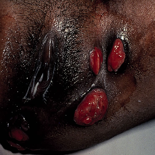
The ulcers may be tender or painless. Secondary infection increases the discomfort. Late in the disease, the patient’s complaints center on scarring and distortion.
The patient’s history usually reveals sexual exposure to a contact in or from an endemic area of India, Africa or the Caribbean.
The lesions may be multiple and can develop in a variety of patterns:
- Nodular with bright red granulating bases
- Hypertrophic with large vegetating masses
- Ulcerative (a combination of the two)
- Cicatricial with spreading scar tissue.
Extragenital lesions occur in the mouth, lips, throat, face, GI tract and bone.3 A degree of lymphedema causes vulvar distortion. There is usually no lymph node involvement.
If left untreated, the areas of involvement may develop squamous cell carcinoma.
Diagnosis
Diagnosis is difficult. Clinicians with cases need to be in touch with the Center for Disease Control www.cdc.gov/mmwr or state laboratory for assistance. Diagnosis requires visualization of dark-staining Donovan bodies of tissue crush preparation or biopsy. In chronic cases, do a biopsy to rule out squamous cell carcinoma.
Pathology/Laboratory Findings
Prepare a tissue smear by removing a fragment of granulation tissue from the base of the ulcer, placing it between two glass slides and staining with Giemsa or Wright’s stain. The Donovan bodies are seen intracellularly and look like safety pins. Cultures are not useful.
Differential diagnosis
Differential diagnosis includes herpes simplex ulcers, syphilitic chancre, chancroid, lymphogranuloma venereum, cutaneous Chron disease, hidradenitis suppurativa, cutaneous tuberculosis, and squamous cell carcinoma.
Treatment and management
CDC recommended regimen4
Doxycycline 100 mg orally twice a day x 3 weeks or until healed
OR
Alternative regimen
Azithromycin 1.0 g orally weekly x 3 weeks or until healed
OR
Ciprofloxacin 750 mg orally twice a day x 3 weeks or until healed
OR
Erythromycin base 500 mg orally four times a day for at least 3 weeks and until all lesions have completely healed
OR
Trimethoprim-sulfamethoxazole DS: 1 tablet (160/800 mg) orally twice a day for at least three weeks or until healed
References
- Fisher BK, Margesson, LJ. Genital Skin Disorders: Diagnosis and Treatment. Mosby, Inc., 1998.146-148.
- Wolff K and Johnson RA. Fitzpatrick’s Color Atlas and Synopsis of Clinical Dermatology, sixth edition. McGraw Hill Medical, 2009. 934.
- Wolff K and Johnson RA. Fitzpatrick’s Color Atlas and Synopsis of Clinical Dermatology, sixth edition. McGraw Hill Medical, 2009. 934.
- Department of health and Human Service, Centers for Disease Control. Sexuallly Transmitted diseases Treatment Guidelines, Morbidity and Mortality Weekly report, 2012;59(RR-12):