Introduction
Mucous membrane pemphigoid (MMP) (also known as cicatricial pemphigoid)1 is a rare, localized, scarring and blistering disease of mucosal surfaces (eyes, mouth, and genitalia) of the elderly.
Epidemiology
Age at onset is between 50 and 70 years. In Western Europe, the incidence is approximately 1-5 cases per million with an age at onset between 60 and 80 years. Women are more commonly affected than men (1.5-2:1).2
Etiology
MMP is an autoimmune condition with autoantibodies directed against the basement membrane zone with resulting inflammation, sub-epidermal splitting with subsequent blistering.
Symptoms and clinical features
Patient history may not be impressive. Patients have involvement most commonly in the mouth (85%), in the eyes (65%), and on the skin (25%). About 50% of patients with MMP have genital disease,3 although an earlier study placed the incidence of genital involvement at 17%.4 There can be itching, vulvar pain, dysuria, and dyspareunia. Purulent vaginal discharge may be present. Patients may have only mild oral or ocular complaints but may progress and develop desquamative gingivitis, conjunctival fibrosis, and scarring.5
The initial vulvar blister may never be seen because it is short-lived. The patient usually presents with erosions involving the mucosal surfaces of the vulva or vagina, eventually progressing to scarring, with loss of the labia and clitoris.
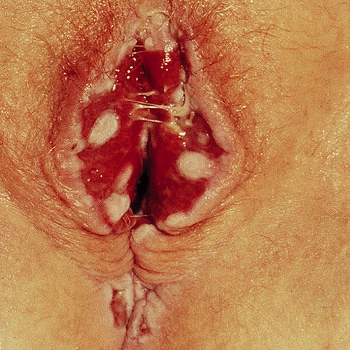
There may be considerable fragility of the vulvar skin, hypo-pigmentation, vaginal erosions, and desquamative vaginitis with copious purulent discharge. Eventual vaginal scarring and obliteration can occur. At the same time, in the mouth, there may be a painful desquamative gingivitis and, in the eyes, considerable scarring and destruction with severe ocular mucosal injury and even blindness. On the skin, scattered tense vesicles or bullae on a red base may rarely occur, usually around the head and neck.
Diagnosis
Diagnosis is essentially clinical. The concomitant oral or ocular symptoms and changes may greatly enhance the ability to diagnose. All mucous membrane diseases that present with erosions must be considered. A biopsy for routine and immunofluorescent histopathology is usually confirmatory.
Pathology/Laboratory Findings
Biopsy is confirmatory, although this autoimmune disorder appears histologically identical to bullous pemphigoid. Immunofluorescent studies can sometimes differentiate between the two. By direct immunoflourescence, 80 to 95% of patients will have autoantibodies directed against the basement membrane zone, with the majority demonstrating linear deposits of IgG and/or C3 on perilesional biopsy specimens.6
Differential diagnosis
Differential diagnosis is similar to that of bullous pemphigoid (pemphigus vulgaris, epidermolyis bullosa acquisita) and also includes erosive lichen planus and lichen sclerosus.
Treatment/management
This can be a difficult, destructive, scarring condition. Suppression of the menses may be needed. Prevention of secondary infection is important.
Management requires the care of a dermatologist working with a dermatopathologist.
General care includes:
- Gentle cleansing with Cetaphil cleanser
- Avoidance of harsh, irritating soaps
- Wearing loose, ventilated clothing
- For painful open areas, compresses or warm or cool sitz baths using solution are recommended to the patient. Also suggested is the use of a bland barrier ointment—plain petrolatum or zinc-oxide ointment.
Specific treatment for erosive and blistering diseases of the vulva requires selecting appropriately from the following:
- High-potency corticosteroid: topical Clobetasol or Halobetasol 0.05% ointment, twice daily; this can also be used with a vaginal applicator for intravaginal involvement (one gram vaginally at bedtime).
- Some clinicians add the anti-inflammatory effects of an antibiotic such as a tetracycline. (Doxycycline or minocycline 100 mg bid orally).7
- Hydrocortisone compounded as a 100mg suppository or 10% in petrolatum; one suppository or one gram of topical inserted vaginally nightly for 14 days, then 2-3 times weekly
- Dapsone 50 to 150 mg daily orally after negative G-6-PD screen
- Prednisone 50 to 100 mg daily orally, depending on severity is helpful in skin disease, but less helpful for mucosal disease.8
- Steroid sparing doses of cyclophosphamide 1 to 2 mg/kg/day orally
- Azathioprine 2 mg/kg/day orally
- Mycophenolate mofetil 2 g/day orally
- Intralesional triamcinolone 5 mg/mL via a 30-gauge needle into small areas after application of topical EMLA anesthetic cream; repeated every 2 to 4 weeks
- Intravenous immunoglobulin has shown promise in the treatment of this disease. The anti-CD20 antibody rituximab, methotrexate, and TNF-a inhibitors such as etanercept may also provide benefit in some patients.9
References
- Fisher BK, Margesson, LJ. Genital Skin Disorders: Diagnosis and Treatment. Mosby, Inc, 1998. 179-180.
- Bernard, P and Borradori, L. “Pemphigoid Group.” Dermatology, edited by Jean Bolognia, Elsevier Limited, 2018.
- Edwards L and Lynch PJ. Genital Dermatology Atlas and Manual, third edition. Wolters Kluwer, 2018. 176-178.
- Ahmed AR, Hombal SM. Cicatricial Pemphigoid. SOInt J Dermatol. 1986 Mar;25(2):90-6.
- Bernard, P and Borradori, L. “Pemphigoid Group.” Dermatology, edited by Jean Bolognia, Elsevier Limited, 2018.
- Bernard, P and Borradori, L. “Pemphigoid Group.” Dermatology, edited by Jean Bolognia, Elsevier Limited, 2018.
- Edwards L and Lynch PJ. Genital Dermatology Atlas and Manual, third edition. Wolters Kluwer, 2018. 176-178.
- Edwards L and Lynch PJ. Genital Dermatology Atlas and Manual, third edition. Wolters Kluwer, 2018. 176-178.
- Bernard, P and Borradori, L. “Pemphigoid Group.” Dermatology, edited by Jean Bolognia, Elsevier Limited, 2018.