Introduction
Bullous pemphigoid1 is the most common of the uncommon autoimmune blistering disorders. The disease results in tense bullae that occur on the lower extremities, the groin, the abdomen, axilla, neck, and forearms in the flexor areas.
Epidemiology
Although it has been reported to occur in childhood, onset is usually after age 60, often later, into the 70’s and 80’s; there is no known racial predilection.
Etiology
The cause is unknown but believed to be an interaction of autoantibody with bullous pemphigoid antigen (BPAG1 and BPAG2, collagen type XVII) in hemidesmosomes of basal keratinocytes followed by complement activation and attraction of neutrophils and eosinophils, in an inflammatory response.2
Symptoms and clinical features
This process causes tissue injury, inflammation, and dermoepidermal separation. The result is a tense, subepidermal blister. There is often a prodromal phase of fixed urticarial plaques, with itching and irritation that can be present for several months. This eruption can be generalized and may be mistaken for persistent Candida or intertrigo. It is located most commonly in the neck, axilla, groin, and the lower part of the abdomen. The blisters later evolve from these plaques. Mucosal sites occur in the mouth in 10-35% of cases3 and, rarely, in the vulvar mucosa causing labial erosions with or without discomfort.
Firm vesicles and tense bullae of various sizes, full of straw-colored fluid, develop on top of the erythematous and indurated plaques of the prodromal phase. The size of the blisters ranges from one to 5 cm. When these blisters break, they leave raw denuded erosions.
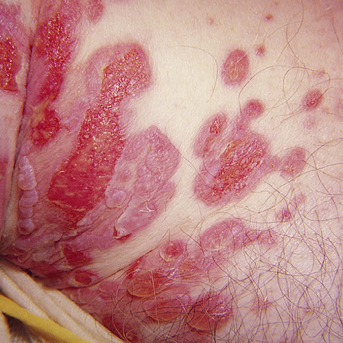
When bullous pemphigoid is localized to the vulva, it is most common on the hairy aspect of the labia majora and inner thighs. Bullous pemphigoid is usually a non-scarring condition, but on occasion it can produce vulvar adhesions. Remission may be spontaneous and complete, or there may be milder recurrences.
Diagnosis
Diagnosis is made on immunofluorescent histopathology ( specimen must be divided with a portion for H&E and a portion in Michel’s medium for immunofluorescent staining), which shows the subepidermal blister with deposition of immunoglobulins along the basement membrane. Circulating anti–basement membrane antibodies can be found in 70% of patients’ sera.
Pathology/Laboratory Findings
Biopsy is necessary to confirm this condition since the levels of cleavage within the superficial skin (subcorneal, intraepidermal, subepidermal, etc.) are diagnostic in bullous disorders.
Differential diagnosis
Differential diagnosis includes pemphigus vulgaris (in which bullae tend to be more flaccid than tense and more likely to affect mucous membranes), bullous drug eruptions (e.g. furosemide, penicillins, psoralens, ibuprofen and some angiotensin-converting enzyme inhibitors)4, cicatricial pemphigoid, erythema multiforme (which usually has more abrupt onset and mucosal lesions), and lichen sclerosus.
Treatment/management
A patient who presents with this picture should be referred to a dermatologist who will, in turn, need to send specific biopsy specimens to a trained dermatopathologist for appropriate diagnosis that will guide management.
Comfort measures such as warm or cool soaks, patting dry, and loose clothing are always useful. A Cochrane Database report published in 2003 noted that, based on review of seven randomized controlled trials, superpotent topical corticosteroids were safe and effective for this condition, even if used long term, and patients had fewer of the side effects associated with oral steroids. In one study, the adjunctive use of steroid-sparing drugs such as azathioprine could reduce the use of oral prednisone. Most of the reported deaths were in patients taking high doses of oral corticosteroids.5
Clinicians with basic skills can start treatment with topical steroids or oral steroids, but it is recommended that patients with extensive disease or those who do not respond to these treatments be referred to a skilled dermatologist.
Treatment of bullous pemphigoid
References
- Fisher BK and Margesson LJ. Genital Skin Disorders: diagnosis and treatment. Mosby, 1998. P. 178-179.
- Wolff K and Johnson RA. Fitzpatrick’s Color Atlas and Synopsis of Clinical Dermatology, sixth addition, McGraw Hill Medical, 2009. 112.
- Wolff K and Johnson RA. Fitzpatrick’s Color Atlas and Synopsis of Clinical Dermatology, sixth addition, McGraw Hill Medical, 2009. 112.
- Edwards L and Lynch P. Genital Dermatology Atlas, second edition, Wolters Kluwer/Lippincott Williams & Wilkins, 2011. 138.
- Khumalo N, Kirtschig G, Middleton P, Hollis S, Wojnarowska F, Murrell D. Interventions for bullous pemphigoid. Cochrane Database Syst Rev. 2003.
- Edwards L and Lynch P. Genital Dermatology Atlas, second edition, Wolters Kluwer/Lippincott Williams & Wilkins, 2011. 138.
- Wolff K and Johnson RA. Fitzpatrick’s Color Atlas and Synopsis of Clinical Dermatology, sixth addition, McGraw Hill Medical, 2009. 113.