Introduction
An angiokeratoma1 is a common, small, harmless, papular, blood vessel tumor with a keratotic surface. A synonym for this condition, when found on the vulva, is angiokeratoma of Fordyce but it should not be confused with Fordyce spots: sebaceous glands sometimes found on the genitalia. The lesion may be solitary or multiple in number. Outside of the benign condition, angiokeratomas may be found in Fabry’s disease or Fucosidosis.
Epidemiology
The prevalence is unknown. In the case of Fabry’s disease, angiokeratomas are present in 66% of males and 36% of females with the disorder.2
Etiology
Their origin is unknown. Patients with multiple angiokeratomas of the vulva, usually with angiokeratomas elsewhere on the body, as well, may have underlying disorders of glycosphingolipid metabolism.
Symptoms and Clinical Features
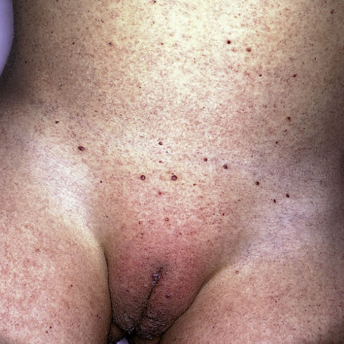
Patient history is asymptomatic vulvar papules. Occasionally a patient may pick at these, causing bleeding. Rarely, mild pruritus may be present.
These common vascular papules are 2 to 5 mm in diameter, range in color from dark red to purplish, and are scattered anywhere on the vulva.
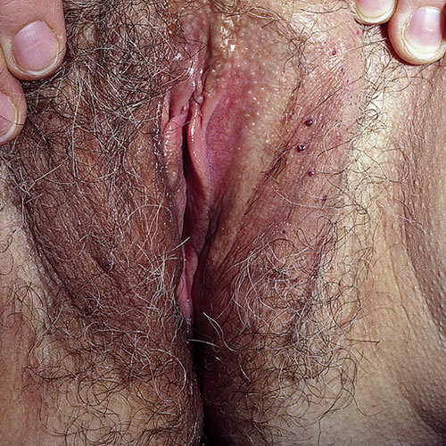
There may be few or sheets of them, and the surface may range from slightly thickened to frankly verrucous. If the lesions are present diffusely outside of the vulva, evaluation for Fabry disease should be undertaken.
Diagnosis
Diagnosis is a combination of clinical and histopathologic. Fabry’s disease (Anderson-Fabry disease or angiokeratoma corporis diffusum) can very rarely present with multiple angiokeratomas of the vulva. There is an X-linked deficiency of alpha-galactosidase-A, resulting in glycosphingolipid accumulation. The condition is mild in women, causing some renal, ocular, and neurologic changes.
Fucosidosis is due to a deficiency of alpha-L-fucosidase resulting in abnormal glycosphingolipid metabolism. Multiple angiokeratomas appear in early childhood on the trunk, groin, upper legs, and in the vulva.
It is associated with severe mental retardation and is lethal.
Pathology/laboratory Findings
Histologically, ectatic (dilated), (and sometimes thrombosed) blood vessels are found in the papillary dermis, along with acanthosis, papillomatosis and prominent epidermal hyperkeratosis, and parakeratosis. 3 4 Biopsy is not usually necessary for the diagnosis.
Differential diagnosis
Differential diagnoses include melanoma, particularly in the case of a single lesion, vulvar warts, and nevi.
Treatment/management
No treatment is needed for asymptomatic lesions. Destruction by electrodesiccation, laser ablation, or occasionally by local excision can be considered.
References
- Fisher BK, Margesson, LJ. Genital Skin Disorders: Diagnosis and Treatment. Mosby, Inc.,1998. 196.
- Zampetti A, Orteu CH, Antuzzi D, et all and the Interdisciplinary group of Fabry disease; article accepted for publication and undergoing revision, 19 Nov 2011. Pubmed.
- Heller, Debra S and Wallach, Robert C, ed. Vulvar Disease: a clinicopathological approach. Informa Healthcare, 2007. 150.
- Cohen PR, Young AW Jr, Tovell HM. Angiokeratoma of the vulva: diagnosis and review of the literature. Obstet Gynecol Surv, 1989 May, 44 (5): 339-346.