Introduction
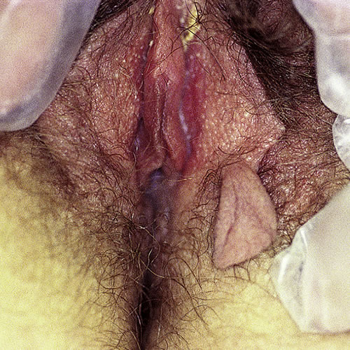
An acrochordon1 is a very common, benign, skin colored, pedunculated, fibroepitheliomatous polyp found in intertriginous areas. Synonyms for acrochordon are skin tag, soft fibroma, papilloma, and fibroepithelial polyp.
Epidemiology
Skin tags occur more often in the middle aged or elderly but may proliferate during pregnancy and then regress postpartum. These lesions, which may be linked to familial tendency, appear in 25% of adults, subsiding in frequency after the age of 502. There is an association between perianal skin tags and Crohn disease, with the skin tags sometimes predating other symptoms of the disease.3
Etiology
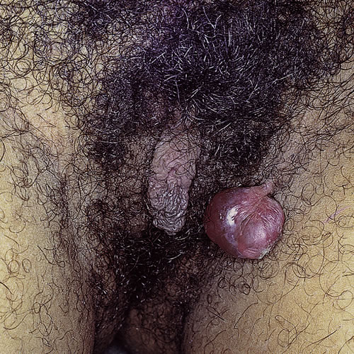
The etiology is unknown. Hormonal or growth factors seem to play a role in obese and diabetic patients. Small acrochorda are referred to as skin tags. The large single lesion is a fibroepithelial polyp.
Symptoms and clinical features
Asymptomatic unless they are traumatized, acrochordons can become inflamed, painful, and can even develop spontaneous necrosis if the pedicle is twisted.

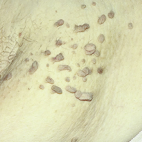
The patient’s usual complaint is that the lesions catch on their clothing. Multiple soft, brown, tan, or skin-colored lesions can be found in the groin and axilla, particularly in patients who are obese and/or diabetic.

Skin tags, (the small ones), may be 2 to 4 mm in diameter and scattered around the sides of the mons pubis and inner thighs. The solitary fibroepithelial polyp is a fleshy, pedunculated mass and may be up to 1.5 cm long on a 2 to 3 mm stalk.

Diagnosis
Diagnosis is clinical.
Pathology/laboratory Findings
Histologically, a thinned epidermis and a loose fibrous tissue stroma is found. 4
Differential diagnosis
Differential diagnoses include pedunculated seborrheic keratosis, nevus, neurofibroma, molluscum, and neuroma.
Treatment/management
Electrodesiccate or snip off the polyps with scissors under local anesthesia. Bleeding can be managed with Monsel’s solution. Asymptomatic lesions do not need treatment.
References
- Fisher BK, Margesson, LJ. Genital Skin Disorders: Diagnosis and Treatment. Mosby, Inc.,1998. 196.
- Banik R and Lubach D. Skin tags: localization and frequencies according to sex and age. Dermatologica, 1987. 174 (4): 180.
- Bonheur JL, Braunstein J, Korelitz B, Panagopoulos G. Anal skin tags in inflammatory bowel disease: new observations and a clinical review. Inflamm Bowel Dis. 2008; 14 (9): 1236.
- Wolff, Klaus and Johnson, Richard A. Fitzpatrick’s Color Atlas and Synopsis of Clinical Dermatology, sixth edition. McGraw Hill Medical. 2009. 231.