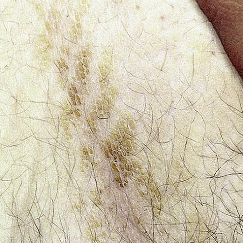
Acanthosis nigricans1(acantho: thorn; nigrican: black) is a non-specific, cutaneous reaction pattern that results in a diffuse, dirty brown, velvety, or warty-surfaced skin change in the axillae and intertriginous body folds, including the inframammary and groin areas.
The benign types are found more commonly in people with naturally darker skin. There is no difference in ethnicity in those who get the form related to malignancy.2 Type I occurs during childhood or puberty. The other types occur when their associated conditions do. Acanthosis has become prevalent in recent years due to the epidemic of obesity.
Acanthosis nigricans includes:
- Type 1: Hereditary benign AN, which starts in childhood and is not associated with endocrinopathy.
- Type 2: Benign (non-malignant) AN associated with endocrine disorders, such as diabetes, acromegaly, Cushing disease, insulin resistance, hirsutism, Addison disease, hypothyroidism. HAIR-AN (hyperandrogenism, insulin resistance, and acanthosis nigricans) syndrome has been described as a common endocrinopathy with distinct pathophysiologic features linking the three conditions together.3
- Type 3: Pseudo-AN, a complication of obesity, related to insulin resistance. Acanthosis nigricans may be an early marker for Type 2 diabetes in both adults and children.45
- Type 4: Drug-induced AN which is caused by nicotinic acid, stilbestrol, niacin, growth hormone treatment, topical fusidic acid and prednisone.6
- Type 5: Malignant AN, part of a paraneoplastic syndrome induced by adenocarcinoma (often of the GI or GU tracts) or lymphoma. In these cases, the tumors themselves are thought to release factors that stimulate keratinocyte proliferation.
Excess growth factor stimulation in the skin produces proliferation of keratinocytes and fibroblasts. These effects may also occur with related conditions.7
Usually there is a gradual onset with no symptoms. The malignant form can be itchy or irritating and of more rapid onset. Hyperpigmentation gradually increases around the neck, in the axillae, in the labiocrural folds, and in the inguinal creases. The skin in these areas becomes thicker and velvety, even warty. In the malignant form, very pronounced pigmentation changes occur and involve the mouth, lips, palms, and soles.
Diagnosis is usually based on the clinical pattern.
There is no specific treatment. If malignancy is suspected, a thorough investigation is indicated. Treatment of the malignancy may cause regression of the acanthosis nigricans.10 Some cases of benign acanthosis nigricans may improve with weight loss and a subsequent change in androgen status or with correction of endocrine disorders. Alphahydroxy acid lotions with 12% lactic acid can be helpful when applied twice a day (Lac-Hydrin lotion). Topical adapalene (Differin) 0.1% gel has also been used with success. These products are best for thinning the thicker lesions and thus decreasing the hyperpigmentation. Topical or systemic retinoids may improve the condition but topical preparations may be irritating to the skin of the crural folds.
References
- Fisher BK and Margesson, LJ. Genital Skin Disorders: Diagnosis and Treatment. Mosby, Inc, 1998. 186.
- Heller DS and Wallach RC, ed. Vulvar Disease: a clinicopathological approach. Informa Healthcare, 2007. 110.
- Barbieri RL, Ryan KJ. Hyperandrogenism, insulin resistance, and acanthosis nigricans syndrome: a common endocrinopathy with distinct pathophysiologic features. Am J Obstet Gynecol. 1983; 147(1):90.
- Keenan BS, Nagamani M. Hyperinsulinema and acanthosis nigricans in African Americans. J Natl Med Assoc. 1997; 89 (8): 523.
- Brickman WJ, Huang J, Silverman BL, et al. Acanthosis nigricans identifies youth at high risk for metabolic abnormalities. J Pediatr. 2010;156:87-92.
- Edwards, Libby and Lynch, Peter. Genital Dermatology Atlas, second edition. Wolters Kluwer;Lippincott Williams and Wilkins, 2011. 228.
- Wolff, Klaus and Johnson, Richard A. Fitzpatrick’s Color Atlas and Synopsis of Clinical Dermatology, sixth edition. McGraw Hill Medical. 2009. 88.
- Heller DS and Wallach RC, ed. Vulvar Disease: a clinicopathological approach. Informa Healthcare, 2007. 110.
- Wolff, Klaus and Johnson, Richard A. Fitzpatrick’s Color Atlas and Synopsis of Clinical Dermatology, sixth edition. McGraw Hill Medical. 2009. 89.
- Wolff, Klaus and Johnson, Richard A. Fitzpatrick’s Color Atlas and Synopsis of Clinical Dermatology, sixth edition. McGraw Hill Medical. 2009. 89.