Introduction
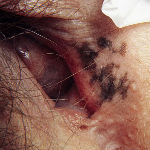
Hyperpigmented macular freckling can occur as a single lesion (lentigo simplex) or as multiple lesions (benign vulvar melanosis). These are completely harmless macules or patches, but they can mimic melanoma.
Epidemiology
Incidence is about 10-15% in women at an average age of 40.
Etiology
The cause is unknown. Melanosis may occur in conjunction with lichen sclerosus.
Symptoms and clinical features
Patients present because of discovery during self-examination or a gynecologic examination.
Irregular brown or black macules or patches with irregular margins scattered anywhere on the vulva—labia majora, minora, or the vestibule.

If the hyperpigmentation is less than 4 mm, it is termed lentigo simplex. Larger lesions, termed vulvar melanosis, can extend to 10 cm or more. Coloration within the lesions can be irregular, the pigmentary changes varying in intensity. The borders can also vary in outline from diffuse to well defined. These patches mostly appear on the labia minora, having a propensity for the less keratinized skin. They may arise in a background of lichen sclerosus.
Diagnosis
Diagnosis is made on histopathology.
Pathology/laboratory findings
Punch biopsy is necessary. On pathological review, there is a clear difference between melanoma and melanosis: “melanosis lacks a substantial melanocytic proliferation, nesting pattern of melanocytes or melanocyte atypia. Dendritic melanocytes are normal in number or only slightly increased along the basal layer of the epidermis in association with hyperpigmentation. Acanthosis may be present.” Lentiginosis is defined by a typical histological pattern.
Differential diagnosis
Because the differential diagnosis includes malignant melanoma and pigmented basal cell carcinoma, as well as post-inflammatory hyperpigmentation, biopsy is strongly recommended.
Lentigines in the vulvar area may be found in some cutaneous syndromes that it is worth being aware of, e.g. Peutz-Jeuger syndrome, LAMB syndrome: Lentigines, atrial myxoma, and blue nevi, and LEOPARD syndrome: Lentigines, electrocardiographic conduction defects, ocular hypertelorism, pulmonary stenosis, abnormalities of the genitalia.
Treatment/management
No treatment is needed of benign melanosis. Biopsy permits informed reassurance to the patient and is the only thing recommended.
References
1 Fisher BK and Margesson LJ. Genital Skin Disorders: diagnosis and treatment. Mosby, 1998. 187.
2 Barnhill RL, Albert LS, Shama SK, et al. Genital lentiginosis: a clinical and histological study. J Am Acad Dermatol. 1990; 22:453-460.
3 Heller DS and Wallach RC. Vulvar Disease: a clinicopathological approach. Informa Healthcare, 2007. 112.